. 2002 Nov;22(22):7769-79.
doi: 10.1128/MCB.22.22.7769-7779.2002.

Oct-1 potentiates CREB-driven cyclin D1 promoter activation via a phospho-CREB- and CREB binding protein-independent mechanism

Affiliations

Oct-1 potentiates CREB-driven cyclin D1 promoter activation via a phospho-CREB- and CREB binding protein-independent mechanism

Séverine Boulon et al. Mol Cell Biol. 2002 Nov.

Abstract

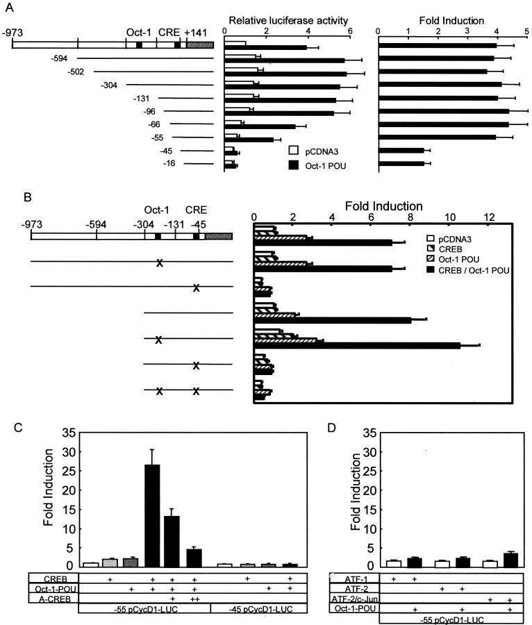
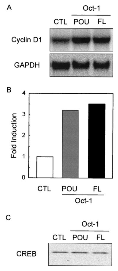
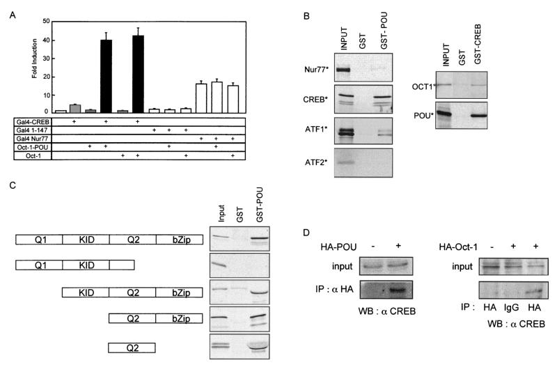
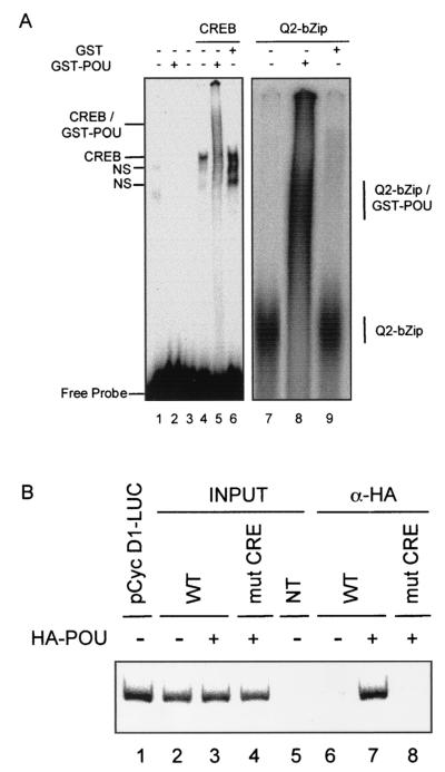
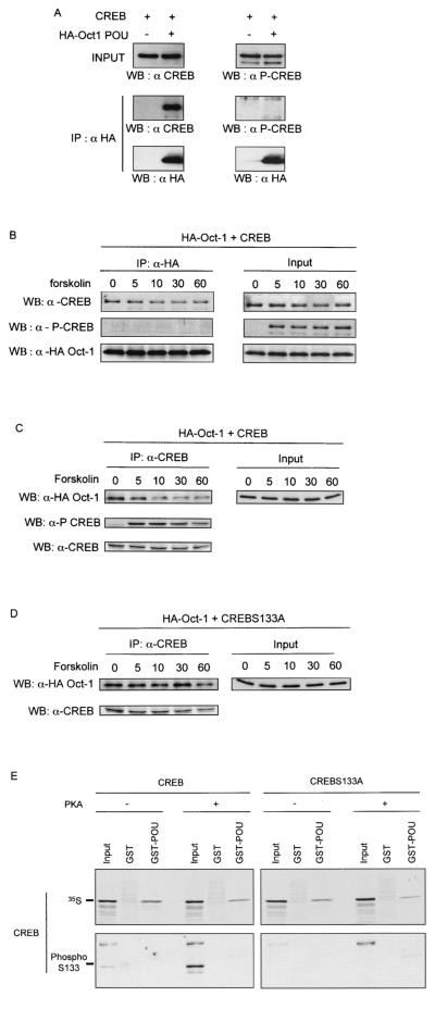
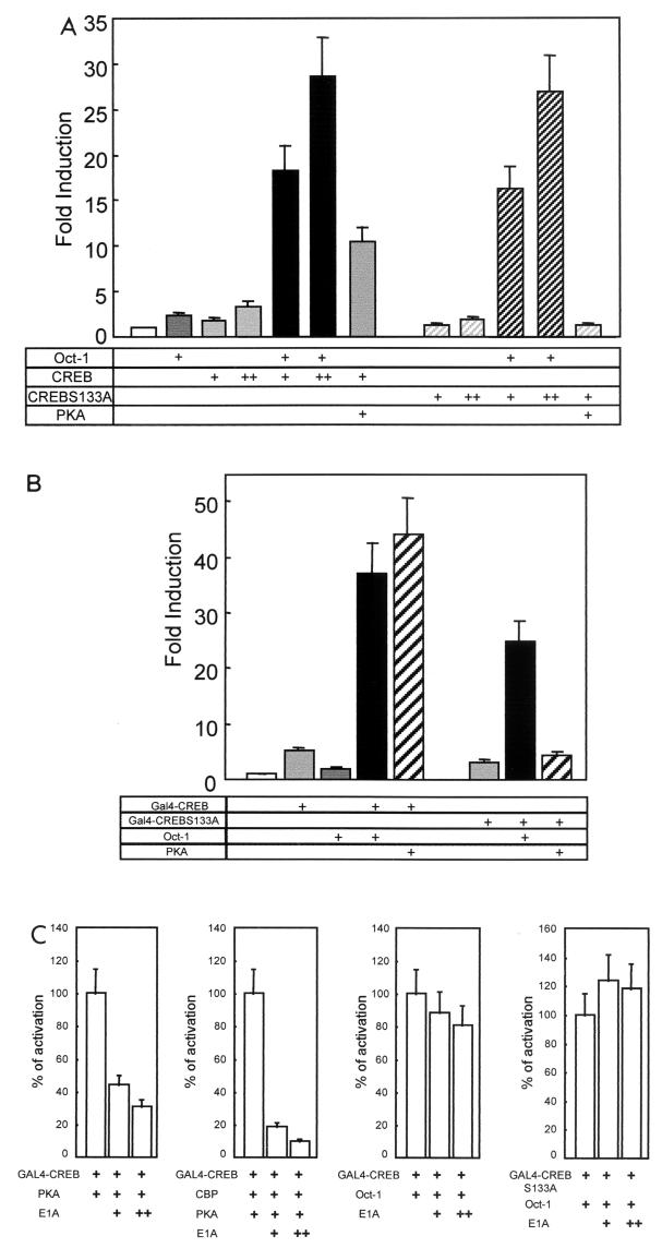
Cyclin D1, the regulatory subunit for mid-G(1) cyclin-dependent kinases, controls the expression of numerous cell cycle genes. A cyclic AMP-responsive element (CRE), located upstream of the cyclin D1 mRNA start site, integrates mitogenic signals that target the CRE-binding factor CREB, which can recruit the transcriptional coactivator CREB-binding protein (CBP). We describe an alternative mechanism for CREB-driven cyclin D1 induction that involves the ubiquitous POU domain protein Oct-1. In the breast cancer cell line MCF-7, overexpression of Oct-1 or its POU domain strongly increases transcriptional activation of cyclin D1 and GAL4 reporter genes that is specifically dependent upon CREB but independent of Oct-1 DNA binding. Gel retardation and chromatin immunoprecipitation assays confirm that POU forms a complex with CREB bound to the cyclin D1 CRE. In solution, CREB interaction with POU requires the CREB Q2 domain and, notably, occurs with CREB that is not phosphorylated on Ser 133. Accordingly, Oct-1 also potently enhances transcriptional activation mediated by a Ser133Ala CREB mutant. Oct-1/CREB synergy is not diminished by the adenovirus E1A 12S protein, a repressor of CBP coactivator function. In contrast, E1A strongly represses CBP-enhanced transactivation by CREB phosphorylated on Ser 133. Our observation that Oct-1 potentiates CREB-dependent cyclin D1 transcriptional activity independently of Ser 133 phosphorylation and E1A-sensitive coactivator function offers a new paradigm for the regulation of cyclin D1 induction by proliferative signals.


Figures

FIG. 1.
FIG. 1.
Oct-1 POU domain-induced cyclin D1 promoter activity is dependent on CREB and the CRE. (A) MCF-7 cells were cotransfected with expression vectors for the POU domain of Oct-1 or empty vector (pCDNA3) with 5′ deletion mutants in the cyclin D1 promoter. The relative luciferase activity was standardized to that observed with pCDNA3 and the full-length promoter. The error bars represent the standard error of the mean. Each experiment was performed at least three times in duplicate (left panel). The right panel shows the induction for each 5′ deletion mutant, calculated by dividing the luciferase activity generated by the POU domain by that observed with pCDNA3 for each deletion mutant. (B) MCF-7 cells were transfected with expression vectors for the POU domain of Oct-1, CREB, or empty vector (pCDNA3), together with a wild-type or mutant cyclin D1-luciferase reporter construct, as indicated schematically to the left. X indicates the mutated site in the promoter. The induction is presented relative to the activity with pCDNA3 and the full promoter, and the error bars represent the standard error of the mean. (C) Cyclin D1 reporters with 5′ deletions to −55 and −45 were transfected with the indicated expression vectors and analyzed as described for B; 100 ng (+) and 300 ng (++) of the dominant negative A-CREB expression vector were cotransfected. (D) Oct-1-POU was coexpressed with ATF-1, ATF-2, and c-Jun as well as the cyclin D1 (−55/+141)-luciferase reporter, as indicated below the bars.
FIG. 2.
FIG. 2.
Oct-1 overexpression increases endogenous cyclin D1 mRNA levels. (A) MCF-7 cells were transfected with either pCDNA3 (CTL), the Oct-1 full-length expression vector (FL), or the POU domain expression vector (POU). RNA was isolated 48 h posttransfection and analyzed by Northern blotting and hybridization with probes for cyclin D1 and GAPDH. (B) The hybridization signals were quantified with a phosphorimager, and the induction was calculated by standardizing to the GAPDH signal. (C) Endogenous CREB protein was detected by immunoblotting with anti-CREB polyclonal antiserum.
FIG. 3.
FIG. 3.
CREB interacts in vitro and in cellulo with Oct-1 independently of DNA binding. (A) MCF-7 cells were transfected as indicated with expression vectors for the GAL4 DNA-binding domain alone (GAL4 1-147), the GAL4 DNA-binding domain fused to CREB (GAL4-CREB) or Nur77 (GAL4-Nur77), full-length Oct-1, the Oct-1 POU domain, or pCDNA3 (first bar). All transfections contained the reporter pG5tk-Luc, which contains five binding sites for GAL4 fused to the thymidine kinase minimal promoter and the firefly luciferase gene. The induction is presented relative to the activity with pCDNA3, and the error bars represent the standard error of the mean. (B) The in vitro-translated, 35S-labeled proteins indicated to the left of each panel were incubated with purified recombinant GST-Oct-1 POU domain (GST-POU), GST-CREB, or GST alone prebound to glutathione-agarose beads. Bound 35S-labeled proteins were visualized by autoradiography of dried SDS-PAGE gels. Input represents 10% of the in vitro-transcribed-translated proteins used in the incubation. (C) The diagram to the left shows the domains of CREB protein present in the in vitro-translated, 35S-labeled proteins that were incubated with GST-POU or GST alone as described for B. Input represents 10% of the in vitro-transcribed-translated proteins. (D) MCF-7 cells were transfected with pCMV-HA-Oct-1 or pCMV-HA-POU, as indicated. The HA fusion proteins were immunoprecipitated (IP) from total cell lysates with anti-HA polyclonal antiserum (HA) or nonspecific rabbit IgG (IgG). Binding of endogenous CREB to the immunoprecipitated protein was detected by immunoblotting (WB) with anti-CREB polyclonal antiserum. Input represents 5% of the total cell lysate.
FIG. 4.
FIG. 4.
Oct-1 POU binds to a CREB-cyclin D1 CRE complex in vitro and in transfected cells. (A) Binding reactions were assembled with a 32P-labeled oligonucleotide spanning the cyclin D1 CRE and the following proteins, as indicated above the lanes: unprogrammed reticulocyte lysate (lane 1); recombinant, full-length CREB (lanes 4 to 6) or the Q2-bZip region of CREB (lanes 7 to 9), produced with programmed reticulocyte lysates; and recombinant GST (lanes 6 and 9) or GST-Oct-1 POU (lanes 2, 5, and 9), purified from bacteria. After electrophoresis on native PAGE, complexes were visualized by autoradiography of the dried gels. The right panel shows the relevant portion of the autoradiogram. Lane 3 shows the probe alone under binding conditions. The composition of the different complexes is indicated to the side of the two panels; NS, nonspecific complexes. (B) Chromatin immunoprecipitation analysis of Oct-1 POU interaction with the cyclin D1 reporter gene in transfected MCF-7 cells. Cells were cotransfected with pCMV-HA-POU (lanes 3, 4, 7, and 8) and either a wild-type −131 pCycD1-LUC reporter gene (WT, lanes 2, 3, 6, and 7) or the same reporter bearing point mutations in the CRE (mut CRE, lanes 4 and 8). After cross-linking, nuclei were purified from transfected cells and sonicated, and after cross-link reversal, an aliquot of chromatin was immunoprecipitated with anti-HA monoclonal antiserum (α-HA, lanes 6 to 8). The cyclin D1 CRE was detected by PCR with primers specific to the cyclin D1-luciferase reporter plasmid in the presence of a trace amount of [α-32P]dCTP. Input indicates PCRs that contained chromatin prior to immunoprecipitation. NT contained chromatin from nontransfected cells (lane 5), while lane 1 shows amplification of the purified plasmid −131 pCycD1-LUC. The panel shows the autoradiogram of the dried gel and is representative of several independent experiments analyzed by 32P incorporation or ethidium bromide staining.
FIG. 5.
FIG. 5.
Oct-1 interacts with unphosphorylated CREB. (A) MCF-7 cells were transfected with pCMV-HA-POU and pCMV-CREB as indicated. HA-Oct-1 POU was immunoprecipitated (IP) from total cell lysates with anti-HA polyclonal antiserum (HA). Binding of CREB and phospho-CREB was detected by immunoblotting (WB) with anti-CREB (α CREB) or anti-P-Ser 133-CREB (α P-CREB) polyclonal antiserum. (B) The same experiment as described for A except that cells were induced with forskolin for the indicated times (in minutes) prior to cell lysis. (C) The same experiment as in B except that the immunoprecipitation (IP) was performed with anti-CREB polyclonal antiserum and Oct-1 binding was revealed by immunoblotting with anti-HA polyclonal antiserum (WB). (D) Identical to C except that anexpression vector for the S133A CREB mutant was transfected. (E) In vitro-translated, 35S-labeled wild-type or S133A mutant CREB was incubated in vitro with ATP alone (−) or together with PKA (+). Subsequently the 35S-labeled proteins were incubated with glutathione-agarose beads bearing either the GST-Oct-1 POU domain fusion protein (GST-POU) or GST alone. In the upper panel, bound 35S-labeled proteins were visualized by autoradiography of dried SDS-PAGE gels. In the lower panel, the binding of phospho-S133A CREB was detected by immunoblotting as described for A. Input represents 10% of the in vitro-transcribed-translated proteins used in the incubation.
FIG.6.
FIG.6.
Oct-1 synergy with CREB is independent of Ser 133 phosphorylation or E1A-sensitive CBP coactivation. (A) MCF-7 cells were transfected with the (−55/+141) cyclin D1-luciferase reporter and, as indicated, with Oct-1, 100 ng (+) or 200 ng (++) of CREB, 100 ng (+) or 200 ng (++) of CREB S133A, or PKA expression vector. The induction is presented relative to the activity with pCDNA3 (bar 1), and the error bars represent the standard error of the mean. (B) MCF-7 cells were transfected with expression vectors for the GAL4 DNA-binding domain fused to wild-type CREB or the S133A mutant, pCMV-Oct-1, and PKA expression plasmid, as indicated. All transfections contained the reporter pG5tk-Luc, which contains five binding sites for GAL4 fused to the thymidine kinase minimal promoter and the firefly luciferase gene. The induction is presented relative to the activity with pCDNA3 (bar 1), and the error bars represent the standard error of the mean. (C) MCF-7 cells were transfected essentially as described (B), except that 150 ng (+) or 300 ng (++) of CBP or E1A expression vectors was included in the transfection, as indicated. The bars represent the luciferase activity as a percentage of that observed in the first lane of each graph, and the error bars present the standard error of the mean.

Similar articles

Cited by

References

    1. Ahn, S., M. Olive, S. Aggarwal, D. Krylov, D. D. Ginty, and C. Vinson. 1998. A dominant-negative inhibitor of CREB reveals that it is a general mediator of stimulus-dependent transcription of c-fos. Mol. Cell. Biol. 18:967-977. - PMC - PubMed
    1. Albanese, C., M. D'Amico, A. T. Reutens, M. Fu, G. Watanabe, R. J. Lee, R. N. Kitsis, B. Henglein, M. Avantaggiati, K. Somasundaram, B. Thimmapaya, and R. G. Pestell. 1999. Activation of the cyclin D1 gene by the E1A-associated protein p300 through AP-1 inhibits cellular apoptosis. J. Biol. Chem. 274:34186-34195. - PubMed
    1. Asahara, H., B. Santoso, E. Guzman, K. Du, P. A. Cole, I. Davidson, and M. Montminy. 2001. Chromatin-dependent cooperativity between constitutive and inducible activation domains in CREB. Mol. Cell. Biol. 21:7892-7900. - PMC - PubMed
    1. Beier, F., R. J. Lee, A. C. Taylor, R. G. Pestell, and P. LuValle. 1999. Identification of the cyclin D1 gene as a target of activating transcription factor 2 in chondrocytes. Proc. Natl. Acad. Sci. USA 96:1433-1438. - PMC - PubMed
    1. Billon, N., D. Carlisi, M. B. Datto, L. A. van Grunsven, A. Watt, X. F. Wang, and B. B. Rudkin. 1999. Cooperation of Sp1 and p300 in the induction of the CDK inhibitor p21WAF1/CIP1 during NGF-mediated neuronal differentiation. Oncogene 18:2872-2882. - PubMed

Publication types

MeSH terms

LinkOut - more resources