. 2002 Nov;22(22):7942-52.
doi: 10.1128/MCB.22.22.7942-7952.2002.

Direct binding of the beta1 adrenergic receptor to the cyclic AMP-dependent guanine nucleotide exchange factor CNrasGEF leads to Ras activation

Affiliations

Direct binding of the beta1 adrenergic receptor to the cyclic AMP-dependent guanine nucleotide exchange factor CNrasGEF leads to Ras activation

Youngshil Pak et al. Mol Cell Biol. 2002 Nov.

Abstract

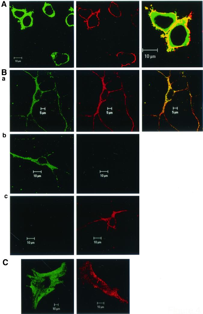
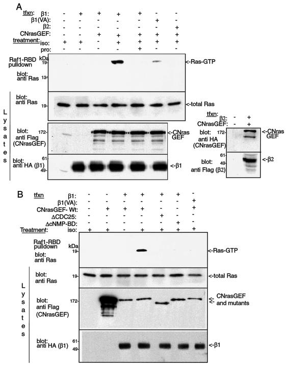
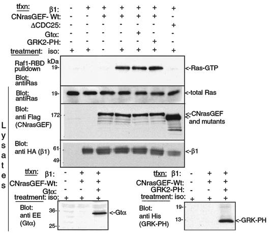
G-protein-coupled receptors (GPCRs) can indirectly activate Ras primarily through the betagamma subunits of G proteins, which recruit c-Src, phosphatidylinositol 3-kinase, and Grb2-SOS. However, a direct interaction between a Ras activator (guanine nucleotide exchange factor [GEF]) and GPCRs that leads to Ras activation has never been demonstrated. We report here a novel mechanism for a direct GPCR-mediated Ras activation. The beta1 adrenergic receptor (beta1-AR) binds to the PDZ domain of the cyclic AMP (cAMP)-dependent Ras exchange factor, CNrasGEF, via its C-terminal SkV motif. In cells heterologously expressing beta1-AR and CNrasGEF, Ras is activated by the beta1-AR agonist isoproterenol, and this activation is abolished in beta1-AR mutants that cannot bind CNrasGEF or in CNrasGEF mutants lacking the catalytic CDC25 domain or cAMP-binding domain. Moreover, the activation is transduced via Gsalpha and not via Gbetagamma. In contrast to beta1-AR, the beta2-AR neither binds CNrasGEF nor activates Ras via CNrasGEF after agonist stimulation. These results suggest a model whereby the physical interaction between the beta1-AR and CNrasGEF facilitates the transduction of Gsalpha-induced cAMP signal into the activation of Ras. The present study provides the first demonstration of direct physical association between a Ras activator and a GPCR, leading to agonist-induced Ras activation


Figures

FIG. 1.
FIG. 1.
β1-AR, but not β2-AR, binds via its C-terminal SkV motif to CNrasGEF.(A) Pulldown assays with the PDZ domain of CNrasGEF. HEK-293T cells were transfected (tfxn) with either HA-tagged wild-type β1-AR (β1) or mutant β1-AR bearing point mutations in the putative PDZ binding motif, SkV (Val→Ala [VA[, Ser→Ala [SA], or Ser →Asp [SD]). Cells were then lysed, and lysates were incubated with either GST alone or a GST fusion protein containing the PDZ domain of CNrasGEF (GST-PDZ). Precipitated proteins were then immunoblotted with anti-HA antibody to detect binding of β1-AR or its mutants. Bottom leftmost lane represents untransfected cell lysate. (B) Same as in panel A, except that β1-AR expressing HEK-293T cells were treated (or not) with a 10 μM concentration of the agonist isoproterenol (Iso) prior to cell lysis and pulldown assays. The phosphatase inhibitor NaF (10 mM) was present during cell treatment and lysis and the pulldown assays. The results shown represent six of seven independent experiments. (C) HEK-293T cells transfected with Flag-tagged β2-AR were lysed as in panel A above, and a pulldown assay performed with GST-PDZ, followed by anti-Flag immunoblotting to detect the β2-AR. No precipitation of the β2-AR was detected despite strong expression of the protein. (D) In the right panel are shown pulldown assay results with GST-PDZ of β1-AR expressed endogenously in primary cultured cortical neurons. Precipitated β1-AR was immunoblotted with anti β1-AR. Ponceau S-stained nitrocellulose shows the total GST or GST-PDZ proteins used in the pulldown experiments in panels A to C. Ten percent of the lysates used for the pulldown experiments in panels A to C are also depicted. The left panel shows parallel immunoblots with anti-β1-AR antibodies and anti-HA antibodies of lysates expressing HA-β1-AR to demonstrate the cleanliness of the anti-β1-AR antibodies used in our studies.
FIG. 2.
FIG. 2.
Direct binding of the PDZ domain of CNrasGEF to the C terminus of β1-AR. Equal amounts (20 μg) of purified His-tagged fusion proteins corresponding to the C terminus of β1-AR (β1-Ct) or the C terminus bearing mutations in the Ser of the SkV motif (β1-Ct [SA] or β1-Ct [SD]) were incubated with GST or GST-PDZ. β1-Ct binding to the GST-PDZ was detected by immunoblotting with anti-His antibodies (upper panel). The lower two panels depict the amounts of the GST/GST-PDZ and His-β1-Ct used for the experiment.
FIG. 3.
FIG. 3.
Coimmunoprecipitation of CNrasGEF and β1-AR coexpressed in HEK-293T cells. Cells expressing HA-tagged β1-AR and Flag-tagged CNrasGEF were lysed, β1-AR immunoprecipitated with anti-HA antibodies and coprecipitated proteins immunoblotted with anti-Flag antibodies. The lower panels depict the expression of the transfected proteins in the cell lysates. The percentage of coimmunoprecipitated proteins relative to total proteins expressed is low.
FIG. 4.
FIG. 4.
Coexpression and colocalization of β1-AR and CNrasGEF. (A) Colocalization of transfected HA-tagged β1-AR (red) and GFP-tagged CNrasGEF (green) in the cell periphery of HEK-293 cells. The β1-AR was detected with anti-HA antibodies. (Ba) Colocalization of endogenous CNrasGEF (green) and β1-AR (red) in primary cultured rat cortical neurons was obtained by double immunostaining with anti-CNrasGEF antibody and anti-β1-AR antibody conjugated with TRITC. (Ba) No β1-AR staining was observed when normal rabbit serum conjugated with TRITC was used as a negative control. (Bb, right panel) Similarly, no CNrasGEF staining was observed when preimmune serum was used as a negative control (Bc, left panel). (C) Endogenous expression of CNrasGEF (green) and β1-AR (red) in coronary artery smooth muscle cells was stained separately.
FIG. 5.
FIG. 5.
Stimulation of β1-AR leads to Ras activation via CNrasGEF in cells. (A) Wild-type β1-AR, but not mutant β1-AR that cannot bind CNrasGEF or β2-AR, activates Ras in the presence of CNrasGEF. HEK-293T cells were transfected with either β1-AR, β1-AR (VA), or β2-AR, along with CNrasGEF, serum starved overnight, and then treated with 10 μM isoproterenol (iso) for 15 min with (+) or without (−) 100 μM propranolol (pro). Cells were then lysed and incubated with immobilized GST-Raf1-RBD (Raf-RBD) to precipitate active (GTP-bound) Ras (11). Ras-GTP was detected with anti-Ras antibodies as shown in the top panel. The lower three panels show the amounts of total Ras (endogenous) and transfected β-adrenergic receptors and CNrasGEF. In the presence of Wt-CNrasGEF, isoproterenol increased Ras activation by 10.9-fold (n = 9). (B) β1-AR-mediated stimulation of Ras requires the presence of intact CNrasGEF. The experiment was performed as in panel A, but mutant CNrasGEF lacking either the catalytic CDC25 domain (ΔCDC25) or the cyclic nucleotide binding (ΔCNMP-BD) domain was expressed together with β1-AR. The data shown are representative of four independent experiments.
FIG. 6.
FIG. 6.
β1-AR stimulation of Ras activation by CNrasGEF is not mediated via Gβγ. (A) HEK-293T cells were transfected with either the transducin α-subunit (EE-tagged Gtα) or the PH domain of GRK2 (His-tagged GRK-PH), along with HA-tagged β1-AR and Flag-tagged CNrasGEF. Cells were treated with 10 μM isoproterenol for 15 min and lysed, and the lysates were then subjected to a Raf-RBD assay to detect active Ras (Ras-GTP, top panel). Expression levels of total (endogenous) Ras and all transfected constructs in cell lysates are depicted in the lower panels.
FIG. 7.
FIG. 7.
β1-AR stimulation of Ras activation by CNrasGEF is transduced via Gsα. (A) Endogenous Gsα can activate Ras via CNrasGEF. HEK-293T cells were transfected with wild-type or mutant CNrasGEF (ΔCDC25 or ΔCNMP-BD) and treated with CTX (30 ng) for 90 min. Cells were then lysed and subjected to Raf-RBD assays. The top panel shows active Ras-GTP. The lower two panels depict the amount of total Ras and the expression of the CNrasGEF proteins. Blots are representative of two independent experiments with virtually identical results. (B) Constitutively active Gsα activates Ras via CNrasGEF, whereas a Gsα inhibitor blocks this activation. HEK-293T cells were transfected with wild-type or mutant CNrasGEF, along with constitutively active Gsα mutants (R201C or Q227L) or an inactive mutant (R232A/I234A), and cells were processed for Raf-RBD assays (top panel) as described above. The R232A/I234A mutation was generated in the context of the R201C mutant. In the presence of Wt-CNrasGEF, constitutively active Gsα mutants increased Ras activation by 2.5- to 3-fold (n = 5) relative to the Gsα mutant alone. The lower panels depict expressed proteins in lysates of the transfected cells and of endogenous total Ras. The blots represent one of five independent experiments.
FIG. 8.
FIG. 8.
Model for β1-AR-mediated Ras activation via CNrasGEF. (A) In the basal state, the β1-AR is associated with the PDZ domain of CNrasGEF via its PDZ binding motif (SkV motif). (B) Agonist binding to the β1-AR results in the dissociation of heterotrimeric G proteins and the activation of Gsα (Gsα-GTP), which in turn activates adenylyl cyclase. Consequently, elevated intracellular cAMP binds to the cNMP-BD of CNrasGEF, activating the catalytic activity of CNrasGEF and leading to Ras activation. (C) After β1-AR stimulation (i.e., after the elevation of cAMP), the serine residue of the SkV motif in the β1-AR carboxyl tail is phosphorylated by GRK2 or GRK5 (GRK), leading to subsequent dissociation of the PDZ domain of CNrasGEF from the β1-AR. For simplicity, only the relevant domains of CNrasGEF (cNMP-BD, PDZ, and CDC25, all in dark gray) are indicated.

Similar articles

Cited by

References

    1. Ambrosini, A., S. Tininini, A. Barassi, G. Racagni, E. Sturani, and R. Zippel. 2000. cAMP cascade leads to Ras activation in cortical neurons. Brain Res. Mol. Brain Res. 75:54-60. - PubMed
    1. Bogoyevitch, M. A., A. J. Ketterman, and P. H. Sugden. 1995. Cellular stresses differentially activate c-Jun N-terminal protein kinases and extracellular signal-regulated protein kinases in cultured ventricular myocytes. J. Biol. Chem. 270:29710-29717. - PubMed
    1. Bos, J. L., J. de Rooij, and K. A. Reedquist. 2001. Rap1 signalling: adhering to new models. Nat. Rev. Mol. Cell. Biol. 2:369-377. - PubMed
    1. Busca, R., P. Abbe, F. Mantoux, E. Aberdam, C. Peyssonnaux, A. Eychene, J. P. Ortonne, and R. Ballotti. 2000. Ras mediates the cAMP-dependent activation of extracellular signal-regulated kinases (ERKs) in melanocytes. EMBO J. 19:2900-2910. - PMC - PubMed
    1. Cass, L. A., S. A. Summers, G. V. Prendergast, J. M. Backer, M. J. Birnbaum, and J. L. Meinkoth. 1999. Protein kinase A-dependent and -independent signaling pathways contribute to cyclic AMP-stimulated proliferation. Mol. Cell. Biol. 19:5882-5891. - PMC - PubMed

Publication types

MeSH terms