. 2002 Nov;22(22):8079-87.
doi: 10.1128/MCB.22.22.8079-8087.2002.

Human Pot1 (protection of telomeres) protein: cytolocalization, gene structure, and alternative splicing

Affiliations

Human Pot1 (protection of telomeres) protein: cytolocalization, gene structure, and alternative splicing

Peter Baumann et al. Mol Cell Biol. 2002 Nov.

Abstract

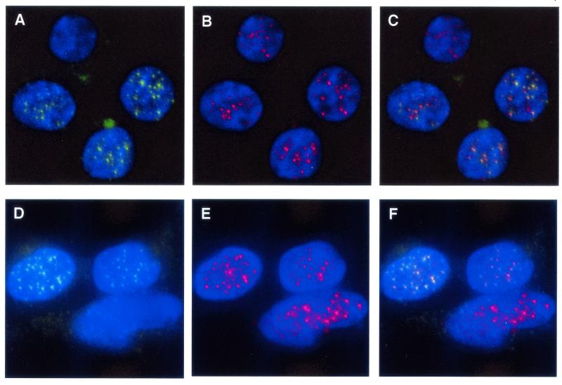
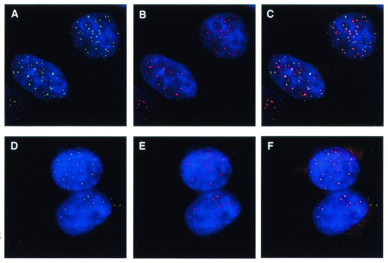
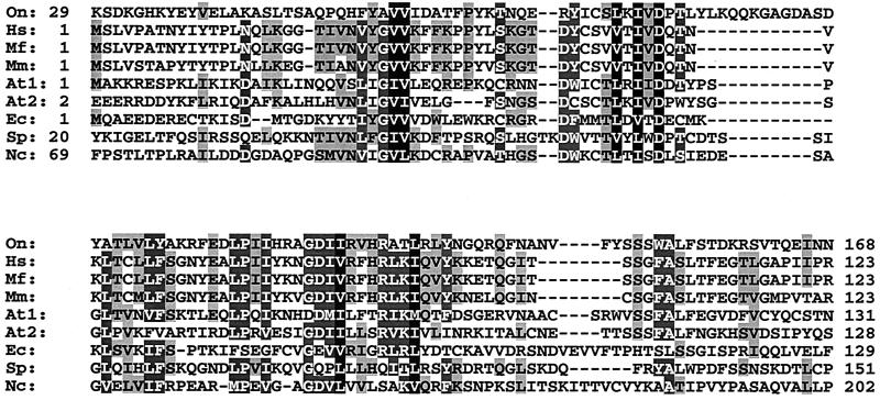
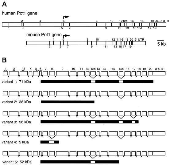
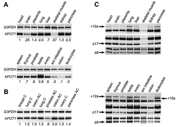
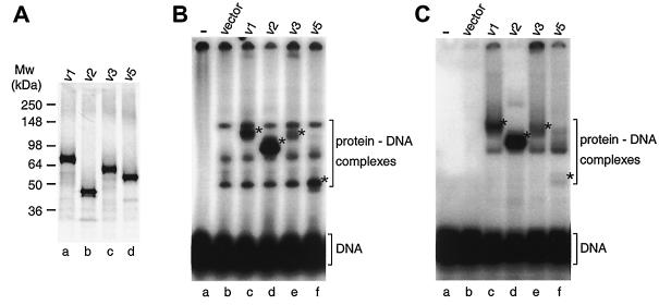
Fission yeast Pot1 (protection of telomeres) is a single-stranded telomeric DNA binding protein with a critical role in ensuring chromosome stability. A putative human homolog (hPot1) was previously identified, based on moderate sequence similarity with fission yeast Pot1 and telomere end-binding proteins from ciliated protozoa. Using indirect immunofluorescence, we show here that epitope-tagged hPot1 localizes to telomeres in interphase nuclei of human cells, consistent with a direct role in telomere end protection. The hPOT1 gene contains 22 exons, most of which are present in all cDNAs examined. However, four exons are subject to exon skipping in some transcripts, giving rise to five splice variants. Four of these are ubiquitously expressed, whereas the fifth appears to be specific to leukocytes. The resultant proteins vary significantly in their ability to form complexes with single-stranded telomeric DNA as judged by electrophoretic mobility shift assays. In addition to these splice variants, the Pot1 family is expanded by the identification of six more genes from diverse species. Pot1-like proteins have now been found in plants, animals, yeasts, and microsporidia.


Figures

FIG. 1.
FIG. 1.
Human Pot1 protein colocalizes with hRap1 in EcR293 cells. Actively growing cells expressing V5-epitope-tagged hPot1 were fixed in formaldehyde and stained for Pot1 with a monoclonal V5 antibody and for hRap1 with a polyclonal rabbit antibody. Nuclei were visualized by 4′,6′-diamidino-2-phenylindole staining of chromosomal DNA. (A and D) Nuclear hPot1 foci (green). Due to the nature of transient transfections, not all cells express V5-tagged Pot1. (B and E) hRap1 staining (red). (C) Merged images from panels A and B; colocalization of V5-hPot1 and hRap1 is indicated by yellow color. (F) Merged images from panels D and E.
FIG. 2.
FIG. 2.
Colocalization of hPot1 with hRap1 and Trf2 in HeLaI.2.11 cells. Actively growing cells expressing V5-epitope-tagged hPot1 were fixed in formaldehyde and stained for Pot1 with a monoclonal V5 antibody and for hRap1 or Trf2 with polyclonal rabbit antibodies. Nuclei were visualized by DAPI staining of chromosomal DNA. (A and D) Nuclear hPot1 foci (green). (B) hRap1 staining (red). (C) Merged images form panels A and B; colocalization of V5-hPot1 and hRap1 is indicated by yellow color. (E) hTrf2 staining (red). (F) Merged images from panels D and E.
FIG. 3.
FIG. 3.
Alignment of N-terminal regions of Pot1-like proteins from Homo sapiens (Hs) (sequence accession number AK001935), M. fascicularis (Mf) (BAB62219), Mus musculus (Mm) (AAH16121), A. thaliana (At1, NP_196249; At2, NP_178592), E. cuniculi (Ec) (CAD26625), S. pombe (Sp) (NP_594453), and N. crassa (Nc) (CAD11392). Protein sequences were aligned in ClustalW using the default values. Amino acids identical or similar in more than four sequences are shown as black letters shaded in gray, amino acids identical or similar in more than six sequences are shown as white letters shaded in gray, and those identical or similar in all sequences are shaded black. The first and last amino acid positions are indicated for each sequence.
FIG. 4.
FIG. 4.
Gene structure and splice variants of hPOT1. (A) Structural organization of human and mouse POT1 genes. Exons are indicated by vertical bars and were numbered after the 20 exons found in the hPOT1 cDNA clone hCT24115 were mapped onto the genomic sequence. The 5′ ends of other hPOT1 cDNA clones are located in exons 2 (accession number AK022580) and 5 (AK001935 and BC002923). Exons specific to splice variants 2 and 5 are numbered 12a and 15a, respectively. Translation of human and mouse POT1 starts in exon 6 and ends in exon 20. (B) Structure of the hPOT1 splice variants. Exons are shown as white boxes (length proportional to exon size); introns are generally long and are not shown to scale. Skipping of an exon is indicated by a bracket below the box. The protein products are shown under each splice variant as black bars. For splice variant 1 the positions of the first 10 AUGs are indicated by asterisks.
FIG. 5.
FIG. 5.
Expression analysis of hPOT1 splice variants. (A) Exons 9 to 12 of hPOT1 were amplified from multitissue cDNA panels, and relative expression levels compared to those for heart (top panel) and spleen (bottom panel) are given. G3PDH was amplified in parallel and used for normalization. (B) Same as panel A except that cDNA samples isolated from tumors were used: breast carcinoma gi-101, lung carcinoma gi-117, colon adenocarcinoma cx-1, lung carcinoma lx-1, prostatic adenocarcinoma pc-3, colon adenocarcinoma gi-112, ovarian carcinoma gi-102, and pancreatic adenocarcinoma gi-103. Breast carcinoma gi-101 was used for normalization. (C) The following regions of hPOT1 mRNA were amplified to detect the presence of different splice forms: exons 12 to 16; products containing exon 12a (variant 2) or exon 15a (variant 5) are indicated by arrows. Exons 16 to 20; product lacking exon 17 (variant 3) is indicated. Exons 6 to 12; product lacking exon 8 (variant 4) is indicated. The unmarked band in each lane corresponds to transcripts that are not variant in this region.
FIG. 6.
FIG. 6.
Binding of hPot1 splice variant proteins to telomeric DNA. (A) sodium dodecyl sulfate-polyacrylamide gel showing in vitro-translated 35S-labeled splice forms 1, 2, 3, and 5. These variants contain 12, 6, 6, and 6 methionine residues, respectively, which accounts for their different signal strengths. Quantification revealed that actual expression levels are within 1.5-fold of each other. (B) Electrophoretic mobility shift assay of a 32P-labeled telomeric oligonucleotide in the presence of hPot1 variants. Protein-DNA complexes specific to hPot1 variants are indicated by asterisks. The three bands seen above the free DNA in lane b represent proteins from the reticulocyte extract binding to the telomeric DNA oligonucleotide and persist in lanes c to f. (C) As with panel B, except that RNA oligonucleotide (UUAGGG)5 was added as competitor.

References

    1. Baumann, P., and T. R. Cech. 2001. Pot1, the putative telomere end-binding protein in fission yeast and humans. Science 292:1171-1175. - PubMed
    1. Bilaud, T., C. Brun, K. Ancelin, C. E. Koering, T. Laroche, and E. Gilson. 1997. Telomeric localization of TRF2, a novel human telobox protein. Nat. Genet. 17:236-239. - PubMed
    1. Blackburn, E. H. 2001. Switching and signaling at the telomere. Cell 106:661-673. - PubMed
    1. Broccoli, D., A. Smogorzewska, L. Chong, and T. de Lange. 1997. Human telomeres contain two distinct Myb-related proteins, TRF1 and TRF2. Nat. Genet. 17:231-235. - PubMed
    1. Cerezo, A., H. Kalthoff, M. Schuermann, B. Schafer, and P. Boukamp. 2002. Dual regulation of telomerase activity through c-Myc-dependent inhibition and alternative splicing of hTERT. J. Cell Sci. 115:1305-1312. - PubMed

Publication types

MeSH terms