. 2002 Oct 29;99(22):14362-7.
doi: 10.1073/pnas.222536599. Epub 2002 Oct 21.

Glycosaminoglycans are a potential cause of rheumatoid arthritis

Affiliations

Glycosaminoglycans are a potential cause of rheumatoid arthritis

Julia Y Wang et al. Proc Natl Acad Sci U S A. .

Abstract

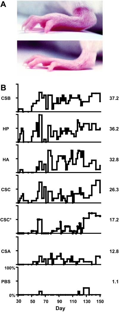
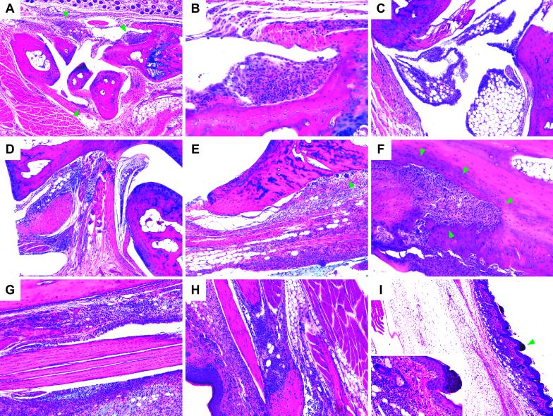
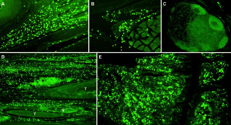
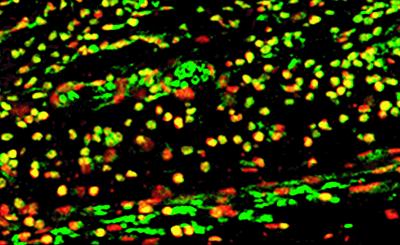
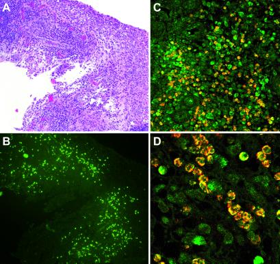
Rheumatoid arthritis (RA) is a chronic, systemic, and inflammatory disease of connective tissue with unknown etiology. We investigated whether aberrant immune responses to glycosaminoglycans (GAGs), a major component of joint cartilage, joint fluid, and other soft connective tissue, causes this disease. Here we show that injection of GAGs such as hyaluronic acid, heparin, and chondroitin sulfates A, B, and C induce arthritis, tendosynovitis, dermatitis, and other pathological conditions in mice. We developed a technique by staining tissue specimens with fluorochrome- or biotin-labeled GAGs to visualize the direct binding between cells and GAGs. We discovered that inflammatory infiltrates from the affected tissue are dominated by a distinct phenotype of GAG-binding cells, a significant portion of which are CD4(+) T cells. GAG-binding cells seem to be expanded in bone marrow of GAG-immunized mice. Furthermore, we identified GAG-binding cells in inflamed synovial tissue of human patients with RA. Our findings suggest that carbohydrate self-antigenic GAGs provoke autoimmune dysfunctions that involve the expansion of GAG-binding cells which migrate to anatomical sites rich in GAGs. These GAG-binding cells might, in turn, promote the inflammation and pathology seen both in our murine model and in human RA.


Figures

Fig 1.
Fig 1.
(A) Examples of a swollen, erythematous rear paw from a mouse treated with CSC (Top) and a normal rear paw from a PBS control mouse (Bottom), both at day 60 of the experiment. Note the involvement of tarsal, metatarsal, and phalangeal joints. (B) Time courses of disease prevalence for mice treated with GAGs or PBS as control. CSC* denotes CSC treatment without Al(OH)3 adjuvant. Ordinates range from 0 to 100% for each graph. Numbers to the right of each graph represent average percentages of diseased mice per day. Note the fluctuating nature of disease progression.
Fig 2.
Fig 2.
(A) Sagittal section through a metacarpus demonstrating global synovial hyperplasia and hypertrophy (focal examples indicated by arrowheads), marked periarthritis, and tendosynovitis. Distal radius to the right; dorsal hair follicles along upper edge. (B) Hyperplastic and hypertrophic (eosinophilic) synovium with lymphoplasmocytic cell infiltration (magnification from A near upper right arrowhead). (C) Hyperplastic and hypertrophic synovia on the dorsal side of an ankle joint. Tibia and calcaneus near upper and right edges, respectively. (D) Pronounced dorsal periarthritis near talocalcaneal and transverse tarsal joints. Talus and calcaneus along lower right and upper edges, respectively. (E) Cell infiltration near distal epiphysis of the tibia with involvement of the extensor tendon sheath. Beginning, pannus-like epiphysial bone erosion involving multinucleate giant cells (arrowhead). (F) Advanced, pannus-like osteo- and chondrolytic lesion in the distal tibia (arrowheads) involving numerous multinucleate giant cells. Marrow cavity in upper right corner. (G) Severe peritendinitis in the tibial extensor compartment. (H) Peritendinitis and dermal cell infiltration near a distal interphalangeal joint. Palmar epidermis to the left. (I) Marked s.c. edema and dermatitis distally in a rear paw. (Inset) Magnification of an area (arrow) with dermal lymphoplasmocytic cell infiltration and parakeratosis. A and B, C and I, and DH are from groups CSC*, HA, and CSC, respectively. Mice from other GAG groups exhibit similar histopathology.
Fig 3.
Fig 3.
GAG staining of tissues from GAG-treated mice demonstrating HA-binding cells. (A) Dermal GAG-binding cell infiltration near a distal interphalangeal joint. Note the epidermis (upper left) and a flexor tendon (bottom). (B) GAG-binding cells in hyperplastic synovium. (C) Scarcity of GAG-binding cells in an activated popliteal lymph node. (D) Connective tissue and tendon sheath infiltration in the extensor compartment near a talocalcaneal joint. T, tendon; V, vein. (E) GAG-binding cells in proximal epiphysial bone marrow of the tibia.
Fig 4.
Fig 4.
Immunostaining showing GAG-binding CD4+ T cells. CD4+ T cells are red, HA-binding cells are green, and costaining is yellow.
Fig 5.
Fig 5.
Left knee synovial tissue specimen from a 33-year-old female patient with RA. (A) Inflamed and hyperplastic synovium with lymphoplasmocytic infiltration (hematoxylin and eosin staining). (B) GAG staining showing HA-binding cell infiltrates (neighboring section from A). (C and D) Costaining for HA-binding CD4+ T cells. CD4+ T cells are red, HA-binding cells are green, and costaining is yellow.

References

    1. Reichlin M. (2001) in Arthritis and Allied Conditions, ed. Koopman, W. (Lippincott, Philadelphia), pp. 1445–1479.
    1. Centers for Disease Control (2001) Morbid. Mortal. Wkly. Rep. 50, 120-125.
    1. Hale L. & Haynes, B. (2001) in Arthritis and Allied Conditions, ed. Koopman, W. (Lippincott, Philadelphia), pp. 1103–1127.
    1. Chakrabarti B. & Park, J. W. (1980) CRC Crit. Rev. Biochem. 8, 225-313. - PubMed
    1. Couchman J. (2001) in Arthritis and Allied Conditions, ed. Koopman, W. (Lippincott, Philadelphia), pp. 209–225.

MeSH terms