

Drusen proteome analysis: an approach to the etiology of age-related macular degeneration
- PMID: 12391305
- PMCID: PMC137479
- DOI: 10.1073/pnas.222551899
Drusen proteome analysis: an approach to the etiology of age-related macular degeneration
Abstract
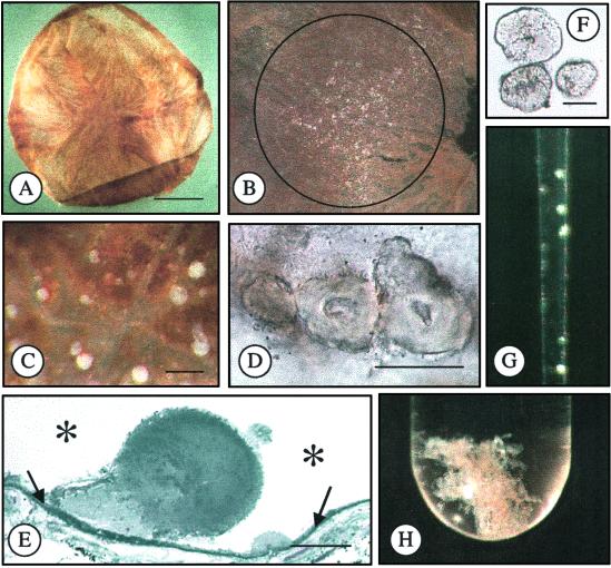
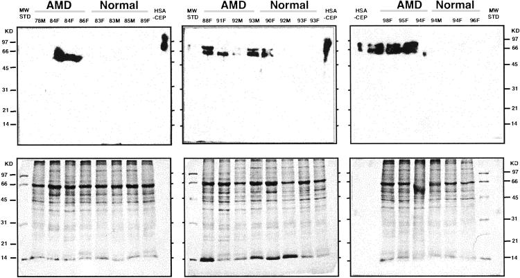
Drusen are extracellular deposits that accumulate below the retinal pigment epithelium on Bruch's membrane and are risk factors for developing age-related macular degeneration (AMD). The progression of AMD might be slowed or halted if the formation of drusen could be modulated. To work toward a molecular understanding of drusen formation, we have developed a method for isolating microgram quantities of drusen and Bruch's membrane for proteome analysis. Liquid chromatography tandem MS analyses of drusen preparations from 18 normal donors and five AMD donors identified 129 proteins. Immunocytochemical studies have thus far localized approximately 16% of these proteins in drusen. Tissue metalloproteinase inhibitor 3, clusterin, vitronectin, and serum albumin were the most common proteins observed in normal donor drusen whereas crystallin was detected more frequently in AMD donor drusen. Up to 65% of the proteins identified were found in drusen from both AMD and normal donors. However, oxidative protein modifications were also observed, including apparent crosslinked species of tissue metalloproteinase inhibitor 3 and vitronectin, and carboxyethyl pyrrole protein adducts. Carboxyethyl pyrrole adducts are uniquely generated from the oxidation of docosahexaenoate-containing lipids. By Western analysis they were found to be more abundant in AMD than in normal Bruch's membrane and were found associated with drusen proteins. Carboxymethyl lysine, another oxidative modification, was also detected in drusen. These data strongly support the hypothesis that oxidative injury contributes to the pathogenesis of AMD and suggest that oxidative protein modifications may have a critical role in drusen formation.
Figures

Comment in
-
New insights and new approaches toward the study of age-related macular degeneration.Proc Natl Acad Sci U S A. 2002 Nov 12;99(23):14619-21. doi: 10.1073/pnas.242607899. Epub 2002 Nov 5. Proc Natl Acad Sci U S A. 2002. PMID: 12419853 Free PMC article. No abstract available.
References
-
- Evans J. R. (2001) Prog. Retinal Eye Res. 20, 227-253. - PubMed
-
- Meyers S. M. & Zachary, A. A. (1988) Arch. Ophthalmol. 106, 651-653. - PubMed
-
- Allikmets R., Singh, N., Sun, H., Shroyer, N. F., Hutchinson, A., Chidambaram, A., Gerard, B., Baird, L., Stauffer, D., Peiffer, A., et al. (1997) Nat. Genet. 15, 236-245. - PubMed
-
- Zhang K., Kniazeva, M., Han, M., Li, W., Yu, Z., Yang, Z., Li, Y., Metzker, M. L., Allikmets, R., Zack, D. J., et al. (2001) Nat. Genet. 27, 89-93. - PubMed
-
- Petrukhin K., Koisti, M. J., Bakall, B., Li, W., Xie, G., Marknell, T., Sandgren, O., Forsman, K., Holmgren, G., Andreasson, S., et al. (1998) Nat. Genet. 19, 241-247. - PubMed
Publication types
MeSH terms
Substances
Grants and funding
LinkOut - more resources
Full Text Sources
Other Literature Sources
Medical
Molecular Biology Databases
