. 2002 Oct 29;99(22):14189-94.
doi: 10.1073/pnas.232310199. Epub 2002 Oct 21.

JLP: A scaffolding protein that tethers JNK/p38MAPK signaling modules and transcription factors

Affiliations

JLP: A scaffolding protein that tethers JNK/p38MAPK signaling modules and transcription factors

Clement M Lee et al. Proc Natl Acad Sci U S A. .

Abstract

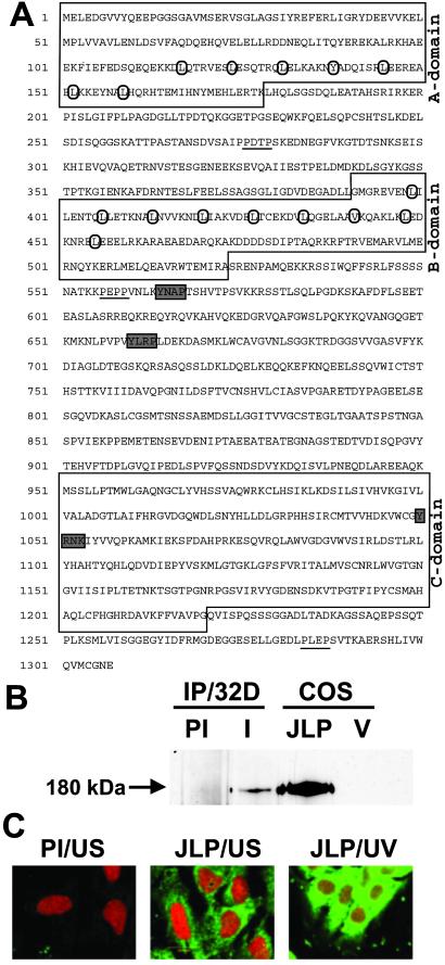
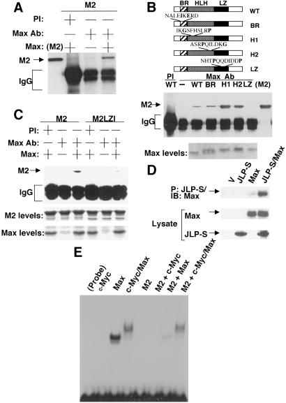
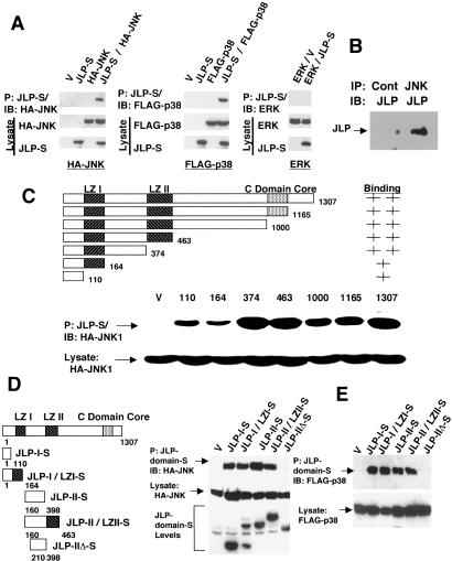
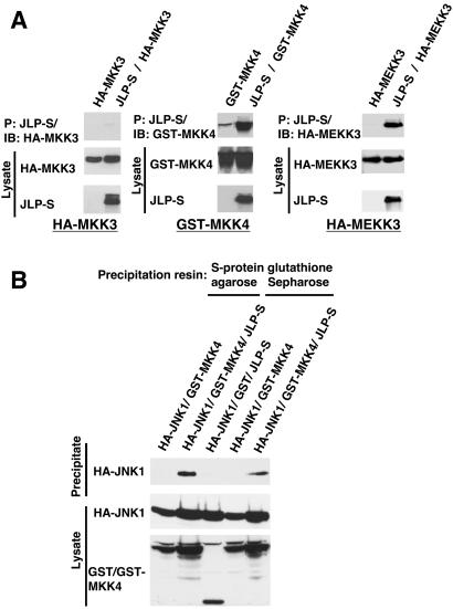
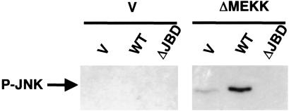
Extracellular signals are transduced into cells through mitogen-activated protein kinases (MAPKs), which are activated by their upstream kinases. Recently, families of scaffolding proteins have been identified to tether specific combinations of these kinases along specific signaling pathways. Here we describe a protein, JLP (c-Jun NH2-terminal kinase-associated leucine zipper protein), which acts as a scaffolding protein to bring together Max and c-Myc along with JNK (c-Jun NH2-terminal kinase) and p38MAPK, as well as their upstream kinases MKK4 (MAPK kinase 4) and MEKK3 (MAPK kinase kinase 3). Thus, JLP defines a family of scaffolding proteins that bring MAPKs and their target transcription factors together for the execution of specific signaling pathways.


Figures

Fig 1.
Fig 1.
Sequence and expression of murine JLP. (A) The deduced protein sequence of JLP is given. The conserved domains A, B, and C are boxed. The heptad repeat of leucine and homologous hydrophobic amino acids in LZI and LZII are circled. The putative SH2 and SH3 binding sites are shaded and underlined, respectively. (B) A full-length cDNA of JLP was subcloned into a modified form of pSG5 mammalian expression vector (Stratagene) and used for transient transfection into COS7 cells. The endogenous JLP of 32Dcl3 cells was immunoprecipitated from cell lysates with preimmune antibody (PI) and the JLP N-terminal-specific antibody (I). The immunoprecipitates were resolved by SDS/PAGE together with the lysates from COS7 cells transfected with vector alone (V) or expression plasmid for JLP. The Western blot analysis was performed with the same JLP antibody. (C) Subcellular localization of JLP. The subcellular localization of endogenous JLP in Swiss3T3 cells was analyzed by indirect immunofluorescence studies. Unstimulated cells (US) were permeablized with Triton X-100, followed by RNase A treatment and immunostaining with preimmune antibody (PI/US) or the JLP C-terminal-specific antibody (JLP/US), followed by the fluorescein-conjugated anti-rabbit IgG secondary antibody (green). The cell nuclei were stained with propidium iodide (red). Cells had also been exposed to UV (254 nm, 5 min), followed by 30-min incubation before they were stained with the JLP-specific antibody (JLP/UV).
Fig 2.
Fig 2.
Association of JLP with Max. (A) Bacterial recombinant Max protein was mixed with the recombinant protein M2 (the N-terminal region of JLP containing LZI and LZII) in the binding buffer (22) and immunoprecipitated with preimmune serum (PI) or Max-specific antibody (Max Ab). The immunocomplexes were subjected to Western blot analysis with the JLP N terminal-specific antibody. (B) A series of bacterial recombinant WT Max and its mutants were used (24). (Top) The mutations were introduced in the basic region (BR), the first helix (H1), the second helix (H2), and the leucine zipper (LZ). The binding studies were carried out by using the method described in A. (Middle) The mapping results from the coimmunoprecipitation experiments. The lane labeled M2 contains the recombinant M2, loaded directly onto the gel. (Bottom) The Coomassie blue-stained gels for the WT and mutants of Max (2 μg each). (C) Mapping of the domain of JLP involved in association with Max. Recombinant WT JLP (M2) or a mutant where the leucines of the first leucine zipper were replaced by alanines (M2LZI) was used in a coimmunoprecipitation experiment with WT Max protein as described in A. The lower two panels show the Coomassie blue-stained gels for the levels of M2, M2LZI, and Max recombinant proteins (5 μg each). (D) In vivo association of JLP with Max. COS7 cells were transiently transfected to express Max with or without S-tagged JLP (JLP-S). The cell lysates were subjected to a pull-down assay using the S-protein agarose. The precipitates as well as the total cell lysates were analyzed with the specific antibodies against the S tag or Max. (E) Effect of JLP on Max homodimerization and c-Myc/Max heterodimerization. Electrophoretic mobility-shift assay was performed by using 32P-labeled double-stranded oligonucleotide, CM1 containing an E-box (CACGTG). Recombinant c-Myc and Max, as described (24), were mixed with the labeled probe in the presence or absence of recombinant M2. The DNA/protein complexes were resolved in a native nondenaturing polyacrylamide gel (4%).
Fig 3.
Fig 3.
Association of JLP with JNK1 and p38MAPKα. (A) The lysates from COS7 cells expressing HA-JNK1 (HA-JNK), FLAG-p38MAPKα (Flag-p38), or endogenous ERK2 with or without JLP-S were precipitated with the S-protein agarose. Cell lysates were prepared as described (26). The precipitates and the lysates were analyzed with the specific antibodies against the S tag, HA tag, FLAG tag, and ERK2. (B) In vivo association of JLP and JNK1. COS7 cells were lysed and immounoprecipitated with JNK1-specific antibody (JNK) or a control rabbit antibody (Cont). The immunoprecipitates were subjected to Western blot analysis with the JLP C terminal-specific antibody (JLP). (C) Deletion mapping of JNK1-interacting domain of JLP. HA-JNK1 was coexpressed with the WT or 3′ deletion mutants of JLP in COS7 cells. All proteins were S-tagged. The lysates were subjected to the pull-down assay with the S-protein agarose. The precipitates and total cell lysates were analyzed with anti-HA antibody. (D) Five mutants of JLP that were C-terminally S-tagged were expressed in COS7 cells along with HA-JNK1. These mutants were JLP-I-S (amino acids 1–110), JLP-I/LZI-S (amino acids 1–164), JLP-II-S (amino acids 160–398), JLP-II/LZII-S (amino acids 160–463), and JLP-IIΔ-S (amino acids 210–398). The cell lysates were precipitated with S-protein agarose, and the precipitates and the total cell lysates were analyzed with the anti-HA antibody on a Western blot. The lower panel shows the expression levels of the JLP mutants detected by using the anti-S-tag antibody. (E) To map the domains of JLP associating with p38MAPKα, pull-down assays using the same set of C-terminally S-tagged proteins of different regions of JLP described in D were used. The proteins were expressed together with FLAG-p38MAPKα in COS7 cells, and the lysates were precipitated with S-protein agarose. The precipitates and the total cell lysates were analyzed with the anti-FLAG tag antibody on a Western blot.
Fig 4.
Fig 4.
In vivo association of JLP with the upstream kinases. (A) COS7 cells were transiently transfected to express the upstream kinases (GST-tagged MKK4, HA-tagged MEKK3, and MKK3) with or without JLP-S. Cell lysates from the transfected cells were subjected to a pull-down assay using the S-protein agarose (26). The precipitates and lysates were analyzed with the corresponding specific antibodies against the S tag, HA tag, or GST tag. (B) To demonstrate ternary complex formation between JLP, JNK1, and MKK4, COS7 cells were transiently transfected to express HA-JNK1 and GST or GST-tagged MKK4 with or without JLP-S as indicated. Cell lysates from the transfected cells were subjected to pull-down assays using the S-protein agarose or glutathione Sepharose. The precipitates and lysates were analyzed with the corresponding specific antibodies against the HA tag or GST tag.
Fig 5.
Fig 5.
Modulation of JNK phosphorylation by JLP. NIH 3T3 cells were transiently transfected with expression plasmids for JNK1-S, FLAG-p38 MAPK, HA-MKK4, and HA-MEKK3 together with JLP-HA WT, its dominant negative mutant containing the deletion of the JNK binding domains (ΔJBD) or an empty vector (V) in the presence or absence of the dominant positive mutant of MEKK1 (ΔMEKK) as indicated. Dominant negative mutant ΔJBD was created by deletion of the amino acids of JLP from 1–107 and 197–209. One day after transfection, cells were lysed and the cell lysates from the transfected cells were subjected to pull-down assays using the S-protein agarose. The precipitates were analyzed with the phospho-specific antibody against JNK1.

Similar articles

Cited by

References

    1. Brunet A. & Pouyssegur, J. (1997) Essays Biochem. 32, 1-16. - PubMed
    1. Cano E. & Mahadevan, L. C. (1995) Trends Biochem. Sci. 20, 117-122. - PubMed
    1. Davis R. J. (1995) Mol. Reprod. Dev. 42, 459-467. - PubMed
    1. Dhanasekaran N. & Reddy, E. P. (1998) Oncogene 17, 1447-1455. - PubMed
    1. English J., Pearson, G., Wilsbacher, J., Swantek, J., Karandikar, M., Xu, S. & Cobb, M. H. (1999) Exp. Cell Res. 253, 255-270. - PubMed

Publication types

MeSH terms

Substances

Associated data