. 2002 Oct 29;99(22):14110-5.
doi: 10.1073/pnas.172527399. Epub 2002 Oct 21.

Pairs of dipeptides synergistically activate the binding of substrate by ubiquitin ligase through dissociation of its autoinhibitory domain

Affiliations

Pairs of dipeptides synergistically activate the binding of substrate by ubiquitin ligase through dissociation of its autoinhibitory domain

Fangyong Du et al. Proc Natl Acad Sci U S A. .

Abstract

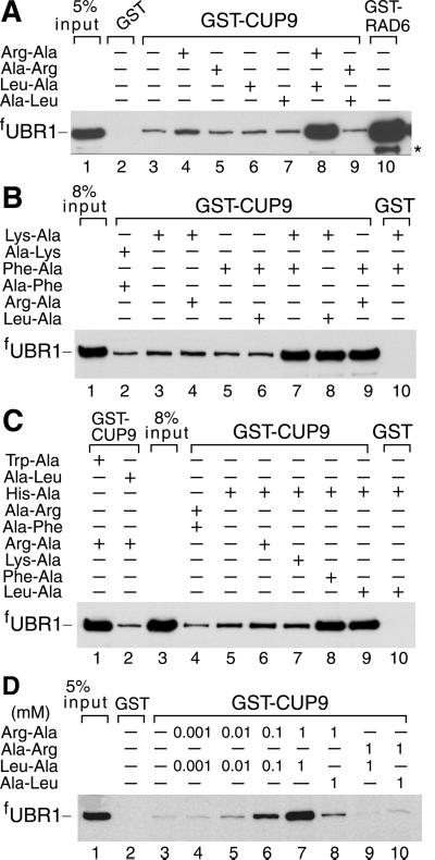
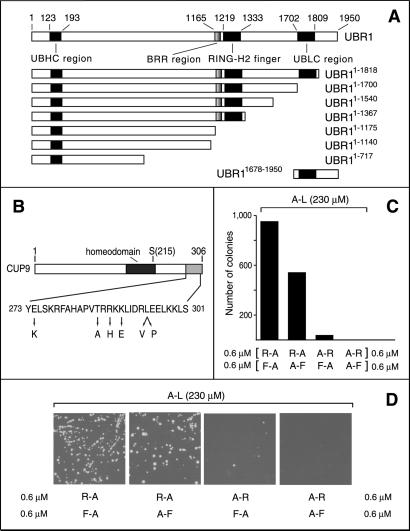
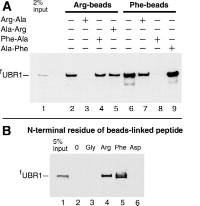
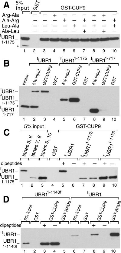
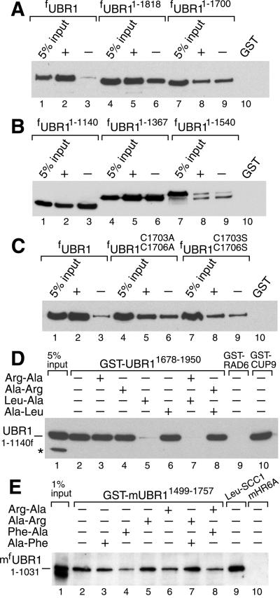
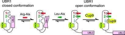
Protein degradation by the ubiquitin (Ub) system controls the concentrations of many regulatory proteins. The degradation signals (degrons) of these proteins are recognized by the system's Ub ligases (complexes of E2 and E3 enzymes). Two substrate-binding sites of UBR1, the E3 of the N-end rule pathway in the yeast Saccharomyces cerevisiae, recognize basic (type 1) and bulky hydrophobic (type 2) N-terminal residues of proteins or short peptides. A third substrate-binding site of UBR1 targets CUP9, a transcriptional repressor of the peptide transporter PTR2, through an internal (non-N-terminal) degron of CUP9. Previous work demonstrated that dipeptides with destabilizing N-terminal residues allosterically activate UBR1, leading to accelerated in vivo degradation of CUP9 and the induction of PTR2 expression. Through this positive feedback, S. cerevisiae can sense the presence of extracellular peptides and react by accelerating their uptake. Here, we show that dipeptides with destabilizing N-terminal residues cause dissociation of the C-terminal autoinhibitory domain of UBR1 from its N-terminal region that contains all three substrate-binding sites. This dissociation, which allows the interaction between UBR1 and CUP9, is strongly increased only if both type 1- and type 2-binding sites of UBR1 are occupied by dipeptides. An aspect of autoinhibition characteristic of yeast UBR1 also was observed with mammalian (mouse) UBR1. The discovery of autoinhibition in Ub ligases of the UBR family indicates that this regulatory mechanism may also control the activity of other Ub ligases.


Figures

Fig 1.
Fig 1.
Type 1 and type 2 dipeptides, if present together, greatly increase the binding of UBR1 to CUP9. Equal amounts of an extract from S. cerevisiae overexpressing the N-terminally flag-tagged UBR1 (fUBR1) were incubated with glutathione-Sepharose beads preloaded with either GST alone, GST-CUP9, or with GST-RAD6, in either the presence or absence of dipeptides. The bound proteins were eluted, fractionated by SDS/PAGE, and immunoblotted with anti-flag antibody. Unless otherwise stated, each dipeptide was present at 1 mM. *, Fragment of fUBR1. The “5% input” lanes refer to a directly loaded sample of the yeast extract that corresponded to 5% of the extract's amount used in the GST-pulldown assays.
Fig 2.
Fig 2.
S. cerevisiae UBR1, CUP9, and plating efficiency assays. (A) A diagram of S. cerevisiae UBR1, indicating the regions conserved in the fungal and metazoan UBR proteins (16, 30). His and Cys are two of the conserved residues in the UBHC region (UBR/His/Cys; residues 123–193) that encompass Gly-173 and Asp-176, which are essential for the integrity of the type 1 substrate-binding site of UBR1 (A. Webster, M. Ghislain, and A.V., unpublished data). See the main text for descriptions of the BRR, RING-H2, and UBLC regions. Also shown are the derivatives of UBR1 used in the GST-CUP9-binding assays and a fragment containing the UBLC domain (used as GST-UBR11678–1950) in the binding assay with UBR11–1140 and UBR11678–1950. UBR1 and its fragments bore N-terminal flag, except for UBR11–1140, which contained C-terminal flag. (B) A diagram of the 306-residue CUP9 highlighting its homeodomain, the site of the toxicity-reducing Asn-215→Ser mutation (13) and the C-terminal region, whose sequence is shown below together with the alterations that decreased the UBR1-dependent degradation of the corresponding CUP9 mutants in yeast (see supporting information). (C) Plating efficiency assays, carried out in minimal medium with S. cerevisiae SC295 (Leu) in the presence of 230 μM Ala-Leu and the indicated pairs of nonnutritious dipeptides at 0.6 μM each. (D) Representative images of plates from the assays in C.
Fig 3.
Fig 3.
Specific binding of the S. cerevisiae ubiquitin ligase UBR1 to peptides bearing destabilizing N-terminal residues. Equal amounts of an extract from S. cerevisiae overexpressing the N-terminally flag-tagged UBR1 (fUBR1) were incubated with microbeads crosslinked to the C terminus of a 12-mer peptide XIFSTDTGPGGC (X = Arg, Phe, Ser, Thr, Gly, Ala, or Asp), followed by washes, elution of the bound proteins, SDS/PAGE, and immunoblotting with anti-flag antibody (see supporting information). (A) Lane 1: 2% of the initial extract's sample. Lanes 2–5: the binding of fUBR1 to beads-linked RIFSTDTGPGGC, bearing N-terminal Arg, in the absence of presence of specific dipeptides. Lanes 6–9: analogous assays with FIFSTDTGPGGC, bearing N-terminal Phe. (B) Lane 1: 5% of the initial extract's sample. Lanes 2–6: fUBR1 binding assay, in the absence of added dipeptides, with either mock-crosslinked beads (0) or the beads with 12-mer peptides bearing N-terminal Gly, Arg, Phe, and Asp, respectively.
Fig 4.
Fig 4.
Dipeptide-independent, high-affinity binding of CUP9 by N-terminal fragments of UBR1. (A) GST-pulldowns with fUBR11–1175 and GST-CUP9 in the presence of different dipeptides (at 1 mM each). (B) As in A, in the presence of 1 mM Arg-Ala and 1 mM Leu-Ala, with either GST alone or GST-CUP9 and either full-length fUBR1, fUBR11–1175, or fUBR11–717. (C) GST-pulldowns with either full-length fUBR1 or fUBR11–1175 in the presence of either 1 mM Arg-Ala and 1 mM Leu-Ala (+) or 1 mM Ala-Arg and 1 mM Ala-Leu (−). Two input amounts of fUBR11–1175, differing by 6-fold, were used (lanes 2 and 3). The corresponding fUBR11–1175 assays are in lanes 7 and 8 vs. 9 and 10, respectively. (D) Comparisons, in the presence of either 1 mM Arg-Ala and 1 mM Leu-Ala (+) or 1 mM Ala-Arg and 1 mM Ala-Leu (−) of the binding of fUBR1 and UBR11–1140f to either GST-CUP9 or GST-RAD6. *, Crossreacting proteins.
Fig 5.
Fig 5.
The C-terminal UBLC domain of UBR1 binds to the N-terminal region of UBR1. (A) GST-pulldowns with GST-CUP9 and either full-length fUBR1, fUBR11–1818, or fUBR11–1700 in the presence of either 1 mM Arg-Ala and 1 mM Leu-Ala (+) or 1 mM Ala-Arg and 1 mM Ala-Leu (−). (B) As in A, but with UBR11–1140f, fUBR11–1367, and fUBR11–1540. (C) As in A, but with full-length WT fUBR1 and its full-length derivatives fUBR1C1703,1706A and fUBR1C1703,1706S. (D) GST-pulldown for the interaction between UBR11–1140f and GST-UBR11678–1950 (see Fig. 2A and the main text) in the presence of different dipeptides. Lanes 9 and 10: results of analogous assays with UBR11–1140f and either GST-RAD6 or GST-CUP9. *, Crossreacting protein. (E) Lanes 1–8: as in D, but with S. cerevisiae extract containing mfUBR11–1031, the N-terminally flag-tagged N-terminal fragment of the mouse UBR1 (E3α) Ub ligase, and with GST-mUBR11499–1757, a C-terminal fragment of mouse UBR1, linked to glutathione-Sepharose beads. Lane 9: GST-pulldown with of mfUBR11–1031 and Leu-SCC1-GST, a GST fusion to a fragment of S. cerevisiae SCC1 (15) bearing N-terminal Leu, a type 2-destabilizing residue. Lane 10: mfUBR11–1031 and GST-mHR6A, one of the cognate mouse E2 enzymes that bind to full-length mUBR1 (16). Lacking the RING-H2 domain, mouse mfUBR11–1031 and yeast UBR11–1140f did not interact with the cognate E2 enzymes, mouse HR6A, and yeast RAD6 (D and E), which bind to full-length mouse and yeast UBR1s, respectively (12, 16). Mouse mfUBR11–1031 retained the substrate-binding sites, as could be demonstrated through its binding to a fragment of the S. cerevisiae SCC1 protein (15) bearing N-terminal Leu, a primary destabilizing residue (lane 9), but not to an otherwise identical SCC1 fragment bearing a stabilizing N-terminal residue (data not shown).
Fig 6.
Fig 6.
Regulated autoinhibition of the ubiquitin ligase UBR1. See the main text for description of the model. The CUP9-binding site of UBR1 is denoted by “i” (a site binding to a substrate's internal degron). The RAD6-binding site is depicted in this orientation solely to indicate its constitutive availability, in contrast to the CUP9-binding site.

References

    1. Hershko A., Ciechanover, A. & Varshavsky, A. (2000) Nat. Med. 10, 1073-1081. - PubMed
    1. Weissman A. M. (2001) Nat. Rev. Mol. Cell. Biol. 2, 169-178. - PubMed
    1. Pickart C. (2001) Annu. Rev. Biochem. 70, 503-533. - PubMed
    1. Conaway R. C., Brower, C. S. & Conaway, J. W. (2002) Science 296, 1254-1258. - PubMed
    1. Zwickl P., Baumeister, W. & Steven, A. (2000) Curr. Opin. Struct. Biol. 10, 242-250. - PubMed

Publication types

MeSH terms

LinkOut - more resources