. 2002 Nov 12;99(23):15060-5.
doi: 10.1073/pnas.212498399. Epub 2002 Oct 21.

The lipid phosphatase myotubularin is essential for skeletal muscle maintenance but not for myogenesis in mice

Affiliations

The lipid phosphatase myotubularin is essential for skeletal muscle maintenance but not for myogenesis in mice

Anna Buj-Bello et al. Proc Natl Acad Sci U S A. .

Abstract

Myotubularin is a ubiquitously expressed phosphatase that acts on phosphatidylinositol 3-monophosphate [PI(3)P], a lipid implicated in intracellular vesicle trafficking and autophagy. It is encoded by the MTM1 gene, which is mutated in X-linked myotubular myopathy (XLMTM), a muscular disorder characterized by generalized hypotonia and muscle weakness at birth leading to early death of most affected males. The disease was proposed to result from an arrest in myogenesis, as the skeletal muscle from patients contains hypotrophic fibers with centrally located nuclei that resemble fetal myotubes. To understand the physiopathological mechanism of XLMTM, we have generated mice lacking myotubularin by homologous recombination. These mice are viable, but their lifespan is severely reduced. They develop a generalized and progressive myopathy starting at around 4 weeks of age, with amyotrophy and accumulation of central nuclei in skeletal muscle fibers leading to death at 6-14 weeks. Contrary to expectations, we show that muscle differentiation in knockout mice occurs normally. We provide evidence that fibers with centralized myonuclei originate mainly from a structural maintenance defect affecting myotubularin-deficient muscle rather than a regenerative process. In addition, we demonstrate, through a conditional gene-targeting approach, that skeletal muscle is the primary target of murine XLMTM pathology. These mutant mice represent animal models for the human disease and will be a valuable tool for understanding the physiological role of myotubularin.


Figures

Fig 1.
Fig 1.
Targeted disruption of mouse Mtm1. (a) The genomic structure of Mtm1 locus surrounding the targeted exon 4, targeting vector, lox-P-flanked Mtm1 exon 4 allele (MTM1L3), and mutant exon 4-deleted allele (MTM1δ4) are shown from top to bottom. Filled and unfilled boxes represent exons and the PGK-Neo cassette, respectively; arrowheads indicate the position of lox-P sequences. The genomic location of probes A, B, and C (long bars) used for Southern blot analysis and PCR primers (short bars) used for screening homologous recombination, exon 4 deletion, and genotyping are also indicated. B, BamHI; Xh, XhoI; P, PstI; Hc, HincII; Xc, XcaI; N, NcoI; H, HindIII; Hc (this site was destroyed after insertion of the PGK-Neo cassette). (b) Mouse genotyping of MTM1L3 line by Southern blot analysis. Genomic DNA from mouse tails (WT, wild-type mouse; HZ, heterozygous female; HM, hemizygous male for the insertion) was digested by BamHI and hybridized with probe A. (c) Mouse genotyping of MTM1δ4 line by PCR amplification. WT and MTM1δ4 alleles result in a 0.9- and 0.2-kb DNA fragment, respectively. KO, knockout hemizygous male. (d) RT-PCR analysis of Mtm1 transcript from quadriceps muscle of either WT or KO mice. Absence of exon 4 in Mtm1 transcript results in a shorter 166-bp DNA fragment in KO sample. (e) Western blot analysis of myotubularin in normal and MTM1δ4 mice. Myotubularin was immunoprecipitated from total protein extracts of quadriceps muscle of wild-type, heterozygous, or KO mice. The antibody used for immunoprecipitation was omitted in the negative control (−).
Fig 2.
Fig 2.
Clinical characterization of Mtm1-deficient mice. (a) The growth rate of KO mice (n = 9) progressively decreases compared with WT males (n = 13). Values were analyzed by ANOVA and show a significant difference (P < 0.0001) between KO and WT animals over time. (b) Clinical evaluation of muscle weakness in mice. (Top and Middle) Illustration of the qualitative tests used to classify mice into different clinical phases. Mutant animals (i and iii) performing either a finger hanging (i and ii) or grid climbing (iii and iv) test, compared with WT mice (ii and iv). (i) Phase II animals are unable to grasp to the finger. (iii) Phase III mice fail in addition to climb on the grid. (v) A phase IV mouse shows signs of kyphosis, because of lesions in paravertebral muscles and hindlimb paralysis. (vi) Illustration of the major muscle mass reduction in a phase IV hindlimb (Left) compared with a normal one (Right). (c) Survival curve of Mtm1-deficient mice (median 52 days, range 37–98, n = 25). (d) Measure of muscular strength by using a dynamometer test. Muscle strength decreases progressively from phase II in KO animals (n = 5 and n = 13 for KO and WT mice, respectively, in phases I and IV, and n = 8 and n = 14 for KO and WT mice, respectively, in phases II and III; **, P = 0.005; ***, P < 0.0001).
Fig 3.
Fig 3.
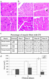
Skeletal muscle histopathology of myotubularin-deficient mice. (a) Hematoxylin and eosin staining of quadriceps cross sections from KO (Upper) and normal (Lower) mice at postnatal day 15 (magnification ×1,000). (b) Mtm1-deficient mice are affected by a generalized centronuclear myopathy. Hematoxylin and eosin staining of phase IV mutant (Upper) and normal (Lower) muscle cross sections from 6-week-old mice (Left, biceps cruralis; Right, triceps brachii). Vacuoles of various sizes in the sarcoplasm of fibers are often present (<, ×630). (c) Percentage of muscle fibers with central or paracentral nuclei in several muscles of mice at different clinical phases (two to seven mice, range 725–3,653 KO and 388–1,558 WT fibers, were analyzed for each point). (d) Muscle fiber area of quadriceps from KO and normal mice. Fiber area is already reduced by 49% in phase II muscle (n = 459 and n = 174 for KO and WT fibers, respectively; *, P < 0.05), and reaches an 83% reduction in phase IV, as compared with normal muscle (n = 891 and n = 538 for KO and WT fibers, respectively; **, P < 0.001).
Fig 4.
Fig 4.
Analysis of myogenic markers in mutant and normal mice. Semiquantitative RT-PCR analysis of myosin heavy chains (embryonic, MHCe; perinatal, MHCp; type I, MHCI; type IIa, MHCIIa; type IIb, MHCIIb; type IIx, MHCIIx), desmin and vimentin transcripts from hindlimb skeletal muscle (supero-posterior compartment, SPC) of wild-type (WT) and KO mice at postnatal day 15 (i) and at 7 weeks of age (phase III, ii). Exon 4 deletion in Mtm1 transcript of KO muscle leads to a smaller PCR product. As negative control (−), the reverse transcriptase was omitted from the sample (n = 2).
Fig 5.
Fig 5.
Structural changes in Mtm1-deficient muscle fibers. (a) Hematoxylin and eosin (iiii) and NADH-TR (ivvi) stainings of muscle cross sections from mutant (i, ii, iv, and v) and normal (iii and vi) hindlimbs of 6-week-old mice (supero-posterior compartment, magnification ×1,000). Note the central (→), peripheral (↓) and anarchical (∗) accumulation of hematoxylin staining and NADH-TR reactivity that labels mitochondria and endoplasmic reticulum, as compared with the homogenous pattern of staining in normal muscle. Occasional fiber splitting (<) can be observed in mutant muscle, as reported in centronuclear myopathy (5). (b) Transmission electron micrographs of hindlimb skeletal muscle (biceps cruralis) from phase IV Mtm1-deficient mice. Longitudinal section of a muscle fiber (Left, 5 weeks) with a centrally located nucleus and perinuclear accumulation of mitochondria, glycogen, and ER is shown (→). Disorganization of myofibrils and sarcomere disarrays is shown. Dilatations of mitochondria in the micrograph are probably artefactual (magnification, ×10,000). Longitudinal section of adjacent myofibers with either normal or degenerative appearance (Right, 13 weeks, ×4,000). Myofibrils are highly disorganized and organelles accumulate in the center of fibers (top).
Fig 6.
Fig 6.
Mtm1 deletion and histopathology in “muscle” (HSA) and “neuronal” (NSE) mutant mice. (a) Mtm1 exon 4 excision in several tissues from conditional KOs. BamHI-digested genomic DNA from brain, heart, quadriceps, and triceps brachii was analyzed by Southern blotting and hybridized with probe C (n = 2 animals for each genotype). (b) Hematoxylin and eosin staining of biceps cruralis cross sections from a 6-week-old HSA (Upper) and an 8-week-old NSE (Lower) mutant mouse.

References

    1. Wallgren-Pettersson C., Clarke, A., Samson, F., Fardeau, M., Dubowitz, V., Moser, H., Grimm, T., Barohn, R. J. & Barth, P. G. (1995) J. Med. Genet. 32, 673-679. - PMC - PubMed
    1. Buj-Bello A., Biancalana, V., Moutou, C., Laporte, J. & Mandel, J. L. (1999) Hum. Mutat. 14, 320-325. - PubMed
    1. Herman G. E., Finegold, M., Zhao, W., de Gouyon, B. & Metzenberg, A. (1999) J. Pediatr. 134, 206-214. - PubMed
    1. Laporte J., Biancalana, V., Tanner, S. M., Kress, W., Schneider, V., Wallgren-Pettersson, C., Herger, F., Buj-Bello, A., Blondeau, F., Liechti-Gallati, S. & Mandel, J. L. (2000) Hum. Mutat. 15, 393-409. - PubMed
    1. Fardeau M., (1992) Skeletal Muscle Pathology (Churchill Livingstone, Edinburgh).

Publication types

MeSH terms

Substances