. 2002 Oct 29;99(22):14206-11.
doi: 10.1073/pnas.212527899. Epub 2002 Oct 21.

Inositol pyrophosphates regulate endocytic trafficking

Affiliations

Inositol pyrophosphates regulate endocytic trafficking

Adolfo Saiardi et al. Proc Natl Acad Sci U S A. .

Abstract

The high energy potential and rapid turnover of the recently discovered inositol pyrophosphates, such as diphosphoinositol-pentakisphosphate and bis-diphosphoinositol-tetrakisphosphate, suggest a dynamic cellular role, but no specific functions have yet been established. Using several yeast mutants with defects in inositol phosphate metabolism, we identify dramatic membrane defects selectively associated with deficient formation of inositol pyrophosphates. We show that this phenotype reflects specific abnormalities in endocytic pathways and not other components of membrane trafficking. Thus, inositol pyrophosphates are major regulators of endocytosis.


Figures

Fig 1.
Fig 1.
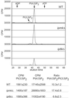
Abnormal accumulation of membranous structures in the absence of inositol pyrophosphates. (Left) HPLC analyses of inositol phosphates in [3H]inositol-labeled yeast by using a PartiSphere SAX column. In ipk1Δ yeast, the Inset represents the HPLC analysis of the putative [PP]2-IP3, using recombinant diphosphoinositol polyphosphate phosphohydrolase protein (DDP1) (12). About 4,000 cpm of the purified 2(PP)-IP3 peak (filled circles) were incubated with 20 ng of recombinant DIPP proteins for 1 h at 37°C (open circles). We observed conversion of 2(PP)-IP3 to PP-IP4, reflecting loss of one of the pyrophosphates, and to IP5, reflecting loss of both pyrophosphate groups. The standards used to identify the inositol phosphates were: IP2, [3H]I(1,4)P2; IP3, [3H]I(1,4,5)P3; IP4, [3H]I(1,3,4,5)P4; IP5, [3H]I(1,3,4,5,6)P5; IP6, [3H]IP6; IP7, [3H]PP-IP5; IP8, [3H]2(PP)-IP4. Transformation of the ipmkΔ and ip6kΔ yeast with the plasmid carrying the deleted genes pIPMK and pIP6K, respectively, restored normal levels of inositol pyrophosphate and normal FM 4-64 internalization. (Right) The lipophilic dye FM 4-64 was used to label yeast membranes for 15 min; cells were then washed and chased for 60 min at 30°C. DIC, dicroic field microscopy. Images were acquired under identical conditions to compare fluorescent intensities.
Fig 2.
Fig 2.
Levels of PI(4,5)P2 do not correlate with aberrant membrane trafficking in ipmkΔ and ip6kΔ yeasts. HPLC analysis of inositol lipids extracted and deacetylated from early logarithmic growth of [3H]inositol-labeled yeast. Deacetylated PI(4,5)P2 was identified by co-elution with authentic standards. The deacetylated PI(3,5)P2 peak was identified by its migration relative to ADP, ATP, and PI(4,5)P2 standards. Data are presented as means ± SE of three or four independent experiments.
Fig 3.
Fig 3.
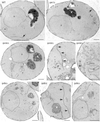
Abnormal membranous structures accumulate in ipmkΔ and ip6kΔ yeasts. Electron microscopy reveals abnormal membranous organelles that accumulate in the absence of inositol pyrophosphate biosynthetic enzymes. The morphology of ipk1Δ yeast is indistinguishable from WT. By contrast, major alterations in ipmkΔ and ip6kΔ yeast are evident. *, stacked membranous cisternae; arrows, ER; #, aberrant organelles; n, nucleus; m, mitochondria; V, vacuole.
Fig 4.
Fig 4.
Aberrant endosomal compartments and processing in cells lacking inositol pyrophosphates. (A) Immunoelectron microscopy with antibodies against the endosomal markers Pep12 and Vph1. Both antibodies specifically recognize membranes accumulated in ipmkΔ and ip6kΔ mutants. (B) Time course of labeling with FM 4-64 in WT and ip6kΔ yeast. After 15 min labeling on ice, cells were washed and incubated at 30°C for 5, 10, or 20 min. In the ip6kΔ mutants after 20 min, the dye is still found in small bright membranous structures. (C) Turnover of the plasma membrane mating pheromone receptor Ste3 (15). The right panel represent a pulse–chase experiment where we observed a retardation of the processing of Ste3 (#) in ip6kΔ yeast compared with WT with an accumulation of a smaller Ste3 fragment (<), reflecting altered processing of the protein and endosomal missorting. (Left) Quantification of three separate experiments. Transformation of the ip6kΔ yeast with the plasmid carrying the deleted gene restored normal Ste3p processing. The asterisks denote significant retardation of Ste3p processing in ip6kΔ yeast (P < 0.05, paired t test).

References

    1. Stephens L., Radenberg, T., Thiel, U., Vogel, G., Khoo, K. H., Dell, A., Jackson, T. R., Hawkins, P. T. & Mayr, G. W. (1993) J. Biol. Chem. 268, 4009-4015. - PubMed
    1. Menniti F. S., Miller, R. N., Putney, J. W., Jr. & Shears, S. B. (1993) J. Biol. Chem. 268, 3850-3856. - PubMed
    1. Shears S. B., Ali, N., Craxton, A. & Bembenek, M. E. (1995) J. Biol. Chem. 270, 10489-10497. - PubMed
    1. Saiardi A., Caffrey, J. J., Snyder, S. H. & Shears, S. B. (2000) J. Biol. Chem. 275, 24686-24692. - PubMed
    1. Timerman A. P., Mayrleitner, M. M., Lukas, T. J., Chadwick, C. C., Saito, A., Watterson, D. M., Schindler, H. & Fleischer, S. (1992) Proc. Natl. Acad. Sci. USA 89, 8976-8980. - PMC - PubMed

Publication types

MeSH terms

Substances

LinkOut - more resources