Review
. 2002 Nov;17(3):143-55.
doi: 10.1002/hbm.10062.

Fast robust automated brain extraction

Affiliations
Review

Fast robust automated brain extraction

Stephen M Smith. Hum Brain Mapp. 2002 Nov.

Abstract

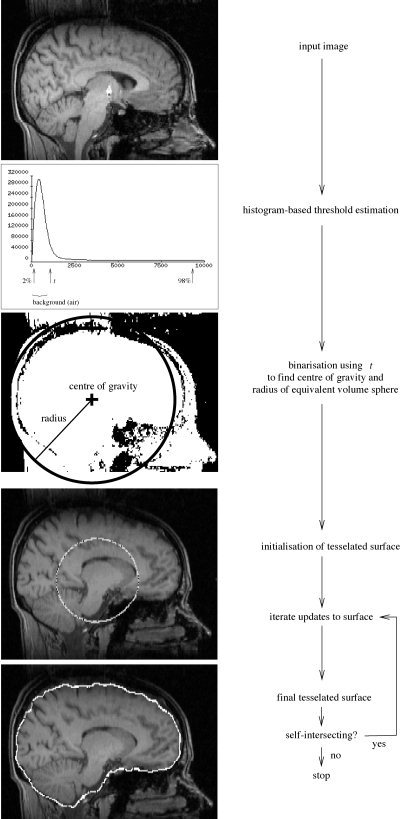
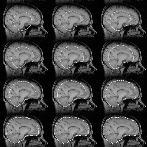
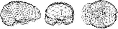
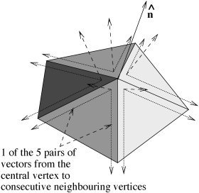
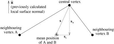
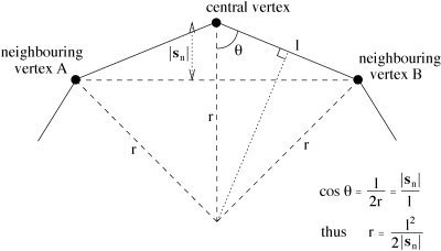
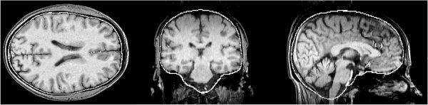
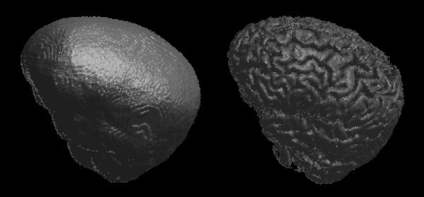
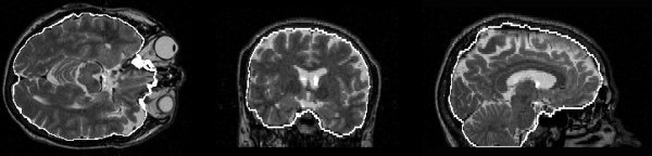
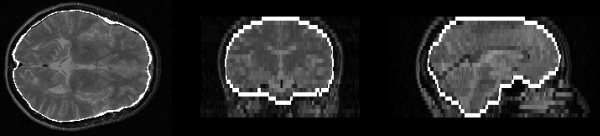
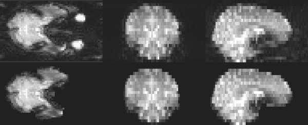
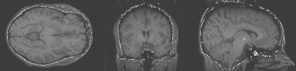
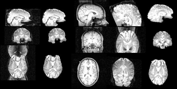
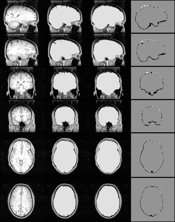
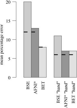
An automated method for segmenting magnetic resonance head images into brain and non-brain has been developed. It is very robust and accurate and has been tested on thousands of data sets from a wide variety of scanners and taken with a wide variety of MR sequences. The method, Brain Extraction Tool (BET), uses a deformable model that evolves to fit the brain's surface by the application of a set of locally adaptive model forces. The method is very fast and requires no preregistration or other pre-processing before being applied. We describe the new method and give examples of results and the results of extensive quantitative testing against "gold-standard" hand segmentations, and two other popular automated methods.


Figures

Figure 1
Figure 1
BET processing flowchart.
Figure 7
Figure 7
Example of surface model development as the main loop iterates. The dark points within the model outline are vertices.
Figure 2
Figure 2
Three views of a typical surface mesh, shown for clarity with reduced mesh density.
Figure 3
Figure 3
Creating local unit vector surface normal from all neighboring vertices.
Figure 4
Figure 4
Decomposing the “perfect smoothness” vector s into components normal and tangential to the local surface.
Figure 5
Figure 5
The relationship between local curvature r, vertex spacing l and the perpendicular component of the difference vector, |s n|.
Figure 6
Figure 6
Smoothness update fraction vs. local radius of curvature, given r max = 10 mm, r min = 3.33 mm.
Figure 8
Figure 8
Example brain surface generated by BET.
Figure 9
Figure 9
Example brain surface model (left) and resulting brain surface (right) generated by BET.
Figure 10
Figure 10
Example brain surface from a T2‐weighted image.
Figure 11
Figure 11
Example brain surface from a proton density image.
Figure 12
Figure 12
Example segmentation of an EPI image.
Figure 13
Figure 13
Example exterior skull surface generated by BET.
Figure 14
Figure 14
Left to right: the original FMRI image; BET output from the FMRI image; T1‐weighted structural image; (failed) registration without using BET; successful registration if BET is used.
Figure 15
Figure 15
Left to right: example original whole‐head MR image; hand segmentation; fully automatic BET masking; hand mask minus BET mask.
Figure 16
Figure 16
Mean % error over 45 MR images for three brain extraction methods, compared to hand segmentation; on the left are the results of testing the fully‐automated methods, on the right are the “hand‐optimized” results. The short bars show the results over only the 35 T1‐weighted images.

References

    1. Atkins M, Mackiewich B (1998): Fully automatic segmentation of the brain in MRI. IEEE Trans Med Imaging 17: 98–107. - PubMed
    1. Bomans M, Höhne K‐H, Tiede U, Riemer M (1990): 3‐D Segmentation of MR images of the head for 3‐D display. IEEE Trans Med Imaging 9: 177–183. - PubMed
    1. Brummer M, Mersereau R, Eisner R, Lewine R (1993): Automatic detection of brain contours in MRI data sets. IEEE Trans Med Imaging 12: 153–166. - PubMed
    1. Cox R. AFNI software . Online: http://afni.nimh.nih.gov.
    1. Dale A, Fischl B, Sereno M (1999): Cortical surface‐based analysis I: segmentation and surface reconstruction. Neuroimage 9: 179–194. - PubMed