. 2002 Nov;3(11):1075-81.
doi: 10.1093/embo-reports/kvf220. Epub 2002 Oct 22.

Expression of mutant Ets protein at the neuromuscular synapse causes alterations in morphology and gene expression

Affiliations

Expression of mutant Ets protein at the neuromuscular synapse causes alterations in morphology and gene expression

Alban de Kerchove D'Exaerde et al. EMBO Rep. 2002 Nov.

Abstract

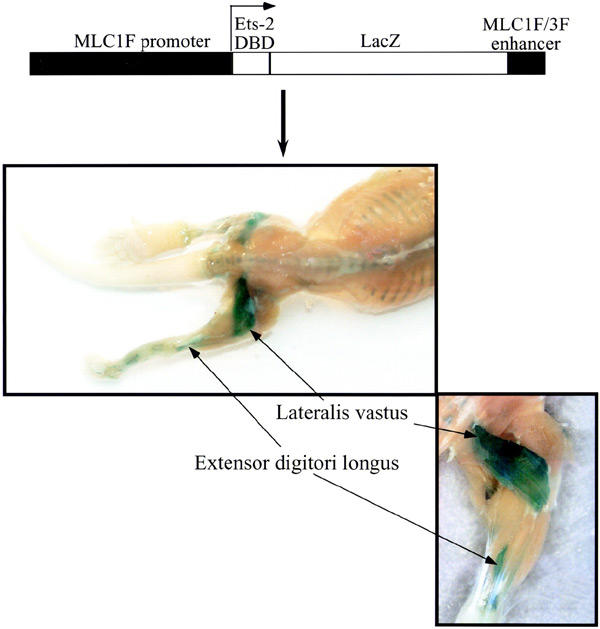
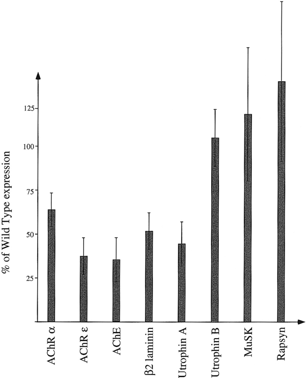
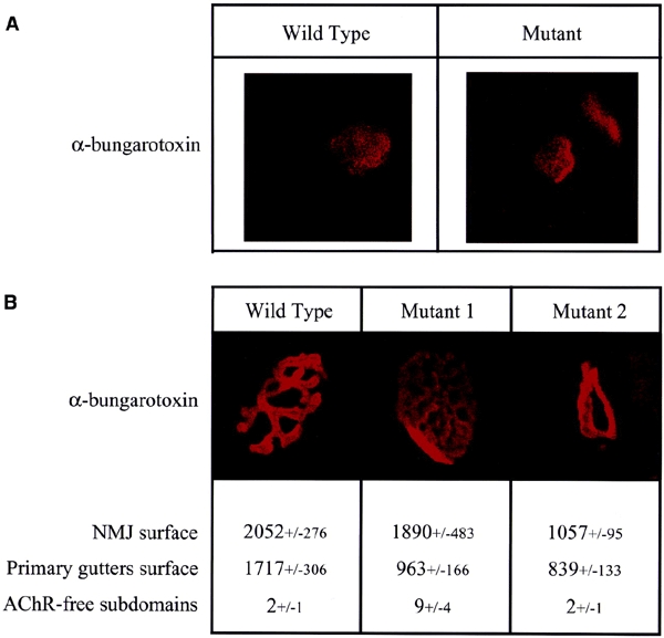
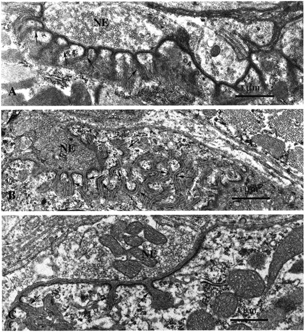
The localized transcription of several muscle genes at the motor endplate is controlled by the Ets transcription factor GABP. To evaluate directly its contribution to the formation of the neuromuscular junction, we generated transgenic mice expressing a general Ets dominant-negative mutant specifically in skeletal muscle. Quantitative RT-PCR analysis demonstrated that the expression of genes containing an Ets-binding site was severely affected in the mutant mice. Conversely, the expression of other synaptic genes, including MuSK and Rapsyn, was unchanged. In these animals, muscles expressing the mutant transcription factor developed normally, but examination of the post-synaptic morphology revealed marked alterations of both the primary gutters and secondary folds of the neuromuscular junction. Our results demonstrate that Ets transcription factors are crucial for the normal formation of the neuromuscular junction. They further show that Ets-independent mechanisms control the synaptic expression of a distinct set of synaptic genes.


Figures

Figure 1
Figure 1
Transgene construction and expression pattern. Transgene expression was detected mainly in the vastus lateralis and extensor digitori longus muscles by in toto LacZ staining of 1-week-old transgenic animals.
Figure 2
Figure 2
Relative expression of synaptic genes in transgenic muscles. The expression levels of the various genes tested are given as percentages of their expression levels in wild-type muscle. Error bars correspond to the standard deviation. Analysis of variance was performed using a one-way ANOVA (P < 0.01 for AChR α and ε, AChE, utrophin A and β2 laminin. Variations of MuSK, Rapsyn and utrophin B expression were not found to be significant). AChR α and ε, acetylcholine receptor subunit gene α and ε; AChE, acetylcholinesterase.
Figure 3
Figure 3
Comparison of the wild-type and mutant NMJ morphologies. NMJs were labelled with rhodaminated α-bungarotoxin and observed by confocal microscopy. (A) One week post-natal NMJ. (B) Three week post-natal NMJ. The NMJ surface corresponds to the surface occupied by the NMJ. Surfaces are expressed in arbitary units. The primary gutter surface corresponds to the surface labelled by, and thus occupied by, the AChR. AChR-free domains correspond to the number of closed surfaces, comprised in the NMJs and not labelled by the α-bungarotoxin. The +/− values correspond to the standard deviation. Mutants 1 and 2 illustrate the two morphological types of mutant NMJ.
Figure 4
Figure 4
Comparative ultrastructural analysis of mutant versus wild-type NMJs. In agreement with confocal studies, the ultrastructural analysis of NMJs of mutant mice on thin sections disclosed severe perturbations in the post-synaptic apparatus. Compared with control animals (A) the post-synaptic membranes of the NMJs in vastus lateralis muscles showed disorganized secondary folds [arrows in (B)]. In other junctions, the folds were scarce or even lacking (C). Nerve endings (NE) looked normal in all muscles. Magnification = 20 000× in (A–C).

References

    1. Agbulut O., Li Z., Perie S., Ludosky M.A., Paulin D., Cartaud J. and Butler-Browne G. (2001) Lack of desmin results in abortive muscle regeneration and modifications in synaptic structure. Cell Motil. Cytoskel., 49, 51–66. - PubMed
    1. Alroy I., Soussan L., Seger R. and Yarden Y. (1999) Neu differentiation factor stimulates phosphorylation and activation of the Sp1 transcription factor. Mol. Cell. Biol., 19, 1961–1972. - PMC - PubMed
    1. Altiok N., Bessereau J.L. and Changeux J.P. (1995) ErbB3 and ErbB2/neu mediate the effect of heregulin on acetylcholine receptor gene expression in muscle: differential expression at the endplate. EMBO J., 14, 4258–4266. - PMC - PubMed
    1. Brandenberger R., Kammerer R.A., Engel J. and Chiquet M. (1996) Native chick laminin-4 containing the β2 chain (s-laminin) promotes motor axon growth. J. Cell Biol., 135, 1583–1592. - PMC - PubMed
    1. Briguet A. and Ruegg M.A. (2000) The Ets transcription factor GABP is required for postsynaptic differentiation in vivo. J. Neurosci., 20, 5989–5996. - PMC - PubMed

Publication types

MeSH terms

Substances

LinkOut - more resources