

Review
doi: 10.1172/JCI16945.
Defective cardiac ion channels: from mutations to clinical syndromes
Affiliations
- PMID: 12393842
- PMCID: PMC150807
- DOI: 10.1172/JCI16945
Item in Clipboard
Review
Defective cardiac ion channels: from mutations to clinical syndromes
J Clin Invest.
2002 Oct.
No abstract available
Figures
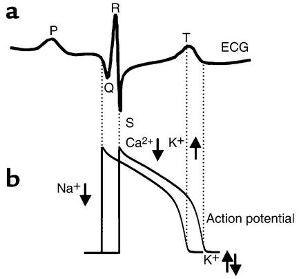
Electrical gradients in the myocardium can be detected on the body surface ECG. (a) An illustrative example of a single cardiac cycle detected as spatial and temporal electrical gradients on the ECG. The P wave is generated by the spread of excitation through the atria. The QRS complex represents ventricular activation and is followed by the T wave reflecting ventricular repolarization gradients. (b) Schematic representation of cellular electrical activity underlying the ECG (see text for details). Where downward arrows represent inward current and upward arrows represent outward current.
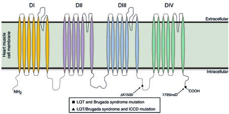
The predicted transmembrane topology of domains I–IV of the cardiac Na+ channel α subunit encoded by SCN5A showing the location and nature of the mutations inducing LQTs, BrS, and isolated cardiac conduction disease.
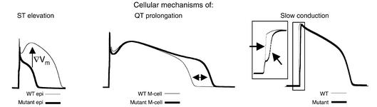
Cellular electrical abnormalities and their relation to changes in the ECG. (Left) At fast pacing (heart) rates, mutation-induced changes in epicardial AP morphologies (thick line) cause dispersion of plateau potentials and a voltage gradient (∇Vm, arrow) (WT, thin line). This gradient will manifest on the ECG as ST segment elevation, indicative of BrS. (Middle) Mutations may prolong APD in M cells (thick line) compared to WT (thin line). The delay in repolarization (ΔAPD ∼ 60ms) is reflected as QT prolongation on the ECG, a hallmark of LQTs. (Right) A rightward shift in the Na+ channel activation curve is sufficient to reduce AP upstroke and slow cardiac impulse conduction (wide QRS segment) as observed in ICCD. WT, wild-type.
Comment on
-
Long QT syndrome, Brugada syndrome, and conduction system disease are linked to a single sodium channel mutation.J Clin Invest. 2002 Oct;110(8):1201-9. doi: 10.1172/JCI15570. J Clin Invest. 2002. PMID: 12393856 Free PMC article.
References
-
- Veldkamp MW, et al. Two distinct congenital arrhythmias evoked by a multidysfunctional Na(+) channel. Circ Res. 2000;86:E91–E97. - PubMed
-
- Rivolta I, et al. Inherited brugada and LQT-3 syndrome mutations of a single residue of the cardiac sodium channel confer distinct channel and clinical phenotypes. J Biol Chem. 2001;276:30623–30630. - PubMed
-
- Bezzina C, et al. A single Na(+) channel mutation causing both long-QT and Brugada syndromes. Circ Res. 1999;85:1206–1213. - PubMed
-
- Tan HL, et al. A sodium-channel mutation causes isolated cardiac conduction disease. Nature (Lond) 2001;409:1043–1047. - PubMed
Publication types
MeSH terms
Substances
LinkOut - more resources
Full Text Sources
Medical
Molecular Biology Databases
