. 2002 Nov;184(22):6235-49.
doi: 10.1128/JB.184.22.6235-6249.2002.

Phenotypes of lexA mutations in Salmonella enterica: evidence for a lethal lexA null phenotype due to the Fels-2 prophage

Affiliations

Phenotypes of lexA mutations in Salmonella enterica: evidence for a lethal lexA null phenotype due to the Fels-2 prophage

Kim Bunny et al. J Bacteriol. 2002 Nov.

Abstract

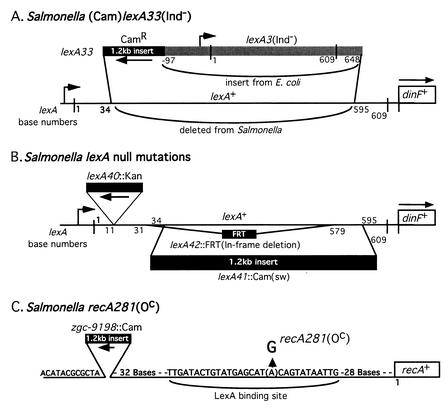
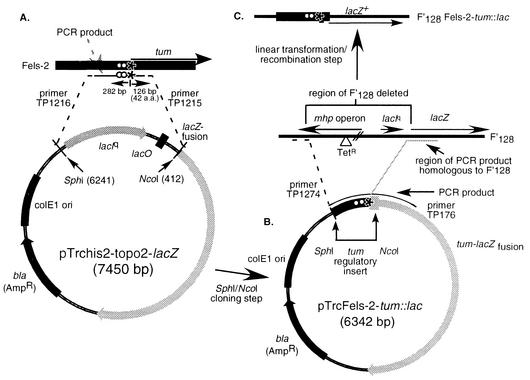
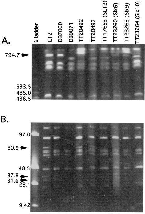
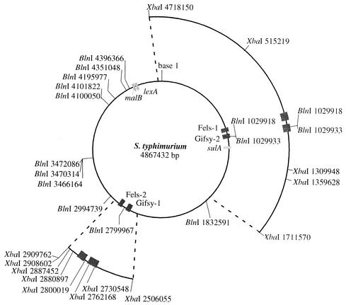
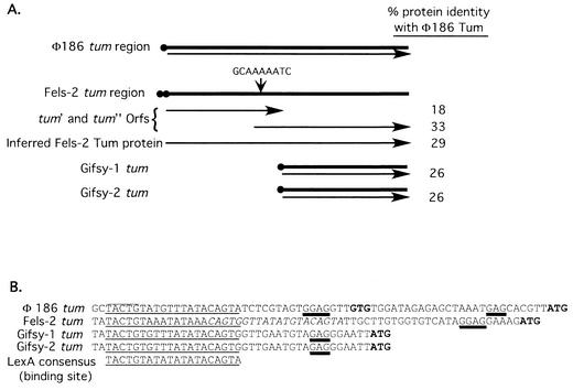
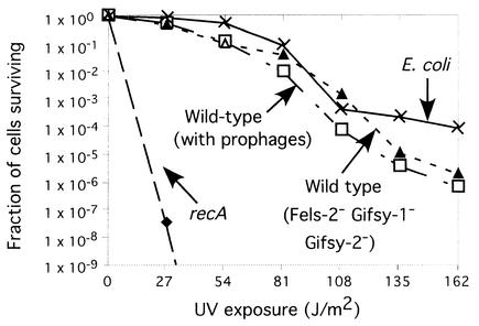
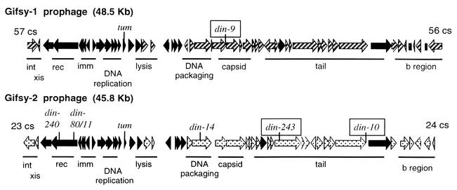
The LexA protein of Escherichia coli represses the damage-inducible SOS regulon, which includes genes for repair of DNA. Surprisingly, lexA null mutations in Salmonella enterica are lethal even with a sulA mutation, which corrects lexA lethality in E. coli. Nine suppressors of lethality isolated in a sulA mutant of S. enterica had lost the Fels-2 prophage, and seven of these (which grew better) had also lost the Gifsy-1 and Gifsy-2 prophages. All three phage genomes included a homologue of the tum gene of coliphage 186, which encodes a LexA-repressed cI antirepressor. The tum homologue of Fels-2 was responsible for lexA lethality and had a LexA-repressed promoter. This basis of lexA lethality was unexpected because the four prophages of S. enterica LT2 are not strongly UV inducible and do not sensitize strains to UV killing. In S. enterica, lexA(Ind(-)) mutants have the same phenotypes as their E. coli counterparts. Although lexA null mutants express their error-prone DinB polymerase constitutively, they are not mutators in either S. enterica or E. coli.


Figures

FIG. 1.
FIG. 1.
Structure of constructed lexA and recA mutations. (A) For the lexA33(Ind) allele, bases 35 to 594 of the Salmonella lexA gene were replaced with a Camr gene and the complete lexA3(Ind) allele of E. coli. (B) The lexA null mutation lexA40::Kan is an insertion of a kanamycin resistance gene after base 11 of lexA and was constructed by Montserrat Llagostera and Xavier Garriga (15). The lexA41::Cam(sw) allele is a replacement of lexA bases 35 to 594 with the above Camr gene. The lexA42::FRT mutation is an in-frame deletion that removes bases 32 to 578. (C) In the recA281(Oc) mutation, a G residue replaces A in the lexA binding site upstream of recA. The Camr gene was inserted upstream to allow selective transduction of this mutation into new strains. Construction methods are described in Materials and Methods.
FIG. 2.
FIG. 2.
Construction of a tum promoter-lacZ reporter construct. (A) The control region of the Fels-2 tum gene was PCR amplified and cloned adjacent to the lacZ gene of the Topo cloning vector (Stratogene). Strains with this high-copy-number plasmid show constitutive lacZ expression. The tum control region was amplified from the above plasmid (B) and inserted (by recombination following linear transformation) adjacent to the lacZ gene of the low-copy-number plasmid F′128 (C). Strains with this plasmid show LexA-controlled expression of LacZ.
FIG. 3.
FIG. 3.
Pulsed-field gel electrophoresis of genomic XbaI fragments from various strains of S. enterica. Arrows indicate bands of interest. For DNA preparation and electrophoresis conditions, see Materials and Methods. (A) High-molecular-weight XbaI bands were separated with pulse times decreasing from 60 to 30 s over a 24-h period. (B) Low-molecular-weight XbaI bands were separated with 7-s pulses over a 24-h period. Strain DB7000 lacks the 90-kb band (due to plasmid pSLT) and also the chromosomal fragments indicated by arrows (see text).
FIG. 4.
FIG. 4.
Genome map of S. enterica, indicating locations of prophages and XbaI and BlnI restriction sites. Boxes represent complete prophage inserted in the Salmonella genome and genes relevant to this study. The published sequence of S. enterica serovar Typhimurium strain LT2 was used to construct this map; sequence coordinates are for a genome that includes all four active prophages (29).
FIG. 5.
FIG. 5.
Comparison of the tum region of coliphage 186 with those from Fels-2, Gifsy-1, and Gifsy-2. (A) Schematic representation of the tum genes of these prophages. Black circles indicate potential LexA binding sites. Arrows indicate the predicted open reading frame(s) for each gene. The potential site for frameshifting in the Fels-2 tum gene is indicated. (B) Potential LexA binding sites (underlined) upstream of the Fels-2, Gifsy-1, and Gifsy-2 tum genes. Sequences are compared to the demonstrated LexA binding region of coliphage 186 tum (8) and the LexA binding site consensus sequence from the review by Walker (56). Italicized bases in the Fels-2 sequence indicate a second potential LexA binding site. Potential start sites for translation are in bold. Predicted Shine-Dalgarno sequences are double underlined. Overlines denote the −10 promoter region identified by Brumby et al. (8).
FIG. 6.
FIG. 6.
UV killing of E. coli and S. enterica. Strains tested were as follows: wild-type S. enterica (recA+ lexA+ sulA+, with all prophages present [TT23656]); S. enterica Fels-2 Gifsy-1 Gifsy-2 (lexA suppressor strain Slx10 made lexA+ and sulA+ [TT23657]); S. enterica recA (recA1 [TT9048]); E. coli K-12 strain, lexA+ sulA+ recA+, without a lambda lysogen [TR7178]).
FIG. 7.
FIG. 7.
Genetic map of the Gifsy phages, with the damage-inducible (din) insertions indicated. Allele numbers point to the site of a din::MudJ insertion. Alleles that are strongly repressed by overexpression of LexA are boxed. Open reading frames in black are nearly identical in both phages, patterned open reading frames show little similarity between Gifsy-1 and Gifsy-2. Nomenclature is as for lambda-like phages: int, integration; xis, excision; red, recombination; imm, immunity; b region, nonessential genes. The positions of the phages in the Salmonella genome are shown in centisomes (cs). (Basic diagram kindly provided by Lionello Bossi and Nara Figueroa.)

References

    1. Akiyoshi, H., and N. Yamamoto. 1970. Genetic evolution of bacteriophage, II. Physical length of the homologous region in a hybrid between serologically and morphologically unrelated phages. Proc. Natl. Acad. Sci. USA 66:385-389. - PMC - PubMed
    1. Anderson, R. P., and J. R. Roth. 1981. Spontaneous tandem genetic duplications in Salmonella typhimurium arise by unequal recombination between ribosomal RNA (rrn) cistrons. Proc. Natl. Acad. Sci. USA 78:3113-3117. - PMC - PubMed
    1. Bagg, A., C. J. Kenyon, and G. C. Walker. 1981. Inducibility of a gene product required for UV and chemical mutagenesis in Escherichia coli. Proc. Natl. Acad. Sci. USA 78:5749-5753. - PMC - PubMed
    1. Benson, N. R., R. M. Wong, and M. McClelland. 2000. Analysis of the SOS response in Salmonella enterica serovar Typhimurium with RNA fingerprinting by arbitrarily primed PCR. J. Bacteriol. 182:3490-3497. - PMC - PubMed
    1. Bergthorsson, U., and H. Ochman. 1995. Heterogeneity of genome sizes among natural isolates of Escherichia coli. J. Bacteriol. 177:5784-5789. - PMC - PubMed

Publication types

MeSH terms

LinkOut - more resources