. 2002 Nov;184(22):6376-83.
doi: 10.1128/JB.184.22.6376-6393.2002.

A proline-rich region with a highly periodic sequence in Streptococcal beta protein adopts the polyproline II structure and is exposed on the bacterial surface

Affiliations

A proline-rich region with a highly periodic sequence in Streptococcal beta protein adopts the polyproline II structure and is exposed on the bacterial surface

Thomas Areschoug et al. J Bacteriol. 2002 Nov.

Abstract

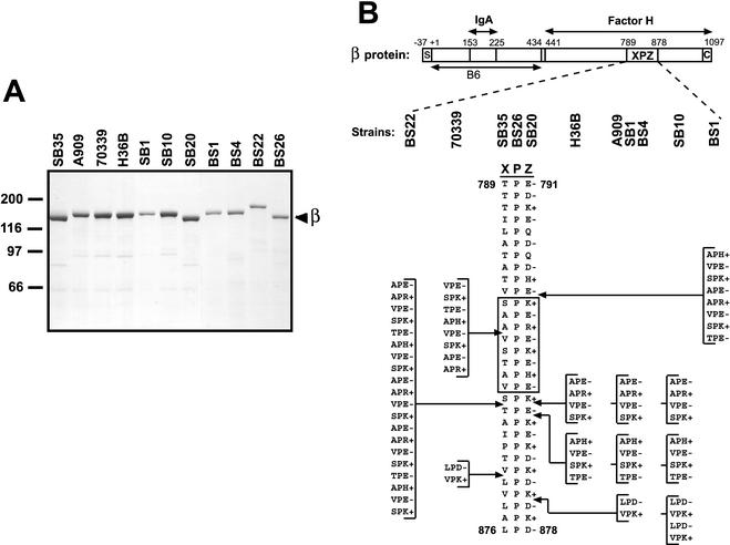
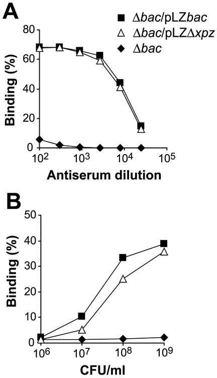
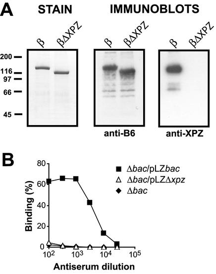
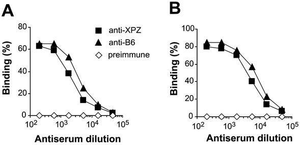
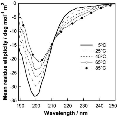
Proline-rich regions have been identified in many surface proteins of pathogenic streptococci and staphylococci. These regions have been suggested to be located in cell wall-spanning domains and/or to be required for surface expression of the protein. Because little is known about these regions, which are found in extensively studied and biologically important surface proteins, we characterized the proline-rich region in one such protein, the beta protein of group B streptococci. The proline-rich region in beta, designated the XPZ region, has a proline at every third position, and the sequence is highly periodic in other respects. Immunochemical analysis showed that the XPZ region was not associated with the cell wall but was exposed on the bacterial surface. Moreover, characterization of a beta mutant lacking the XPZ region demonstrated that this region was not required for surface expression of the beta protein. Comparison of the XPZ region in different beta proteins showed that it varied in size but always retained the typical sequence periodicity. Circular dichroism spectroscopy indicated that the XPZ region had the structure of a polyproline II helix, an extended and solvent-exposed structure with exactly three residues per turn. Because of the three-residue sequence periodicity in the XPZ region, it is expected to be amphipathic and to have distinct nonpolar and polar surfaces. This study identified a proline-rich structure with unique properties that is exposed on the surface of an important human pathogen.


Figures

FIG. 1.
FIG. 1.
Size and sequence variability in the XPZ region of the β protein. (A) Analysis by SDS-PAGE of β proteins expressed by 11 different GBS strains. These β proteins were isolated by incubating washed bacteria at elevated pH, a procedure that allows recovery of almost pure β protein (35). The major band at ∼125 kDa represents the β protein (see text). (B) Schematic representation of the streptococcal β protein and of the proline-rich XPZ region. Binding sites for human IgA-Fc and factor H are located in the N- and C-terminal halves of β, respectively, as indicated. The XPZ region, which is located in the C-terminal part of the protein, is composed of tandemly arranged three-residue XPZ motifs, in which the first residue (X) is uncharged, the second residue (P) is a proline, and the third residue (Z) is almost invariably charged. Moreover, the third residue is alternately of positive and negative charge, as indicated. The XPZ region is not required for binding of factor H but may enhance binding of this ligand (; T. Areschoug, unpublished data). The region designated B6 represents an N-terminal fragment of β used to make antiserum (see text). S, signal peptide; C, C-terminal membrane anchor. The numbers refer to amino acid positions in the processed protein, with numbering according to Hedén et al. (19). The lower part of the figure shows the sequence of the XPZ region in the 11 different β proteins analyzed. Strain designations are indicated at the top. The different XPZ sequences may all be derived from the shortest sequence by the addition of blocks containing multiples of six amino acid residues, as indicated. For example, the amino acid sequence in strain SB10 can be derived from that in strain SB35 by the addition of three 12-residue blocks; the first two of these blocks are the same as those indicated for strain H36B. Due to the periodic sequence of the XPZ region at the protein and DNA levels, other sites of insertion are also possible. The boxed region corresponds to a synthetic peptide used for CD analysis and for raising antiserum to the XPZ region.
FIG. 2.
FIG. 2.
A mutant of the β protein lacking the XPZ region is expressed on the bacterial surface. (A) Surface expression of β was analyzed in the Δbac strain, in which the chromosomal bac gene (encoding β) has been deleted, in the Δbac strain transcomplemented with a plasmid carrying the intact bac gene (Δbac/pLZbac), and in the Δbac strain transcomplemented with a plasmid encoding a β protein (βΔXPZ) lacking the XPZ region (Δbac/pLZΔxpz). The analysis was performed with an antiserum directed against the N-terminal B6 fragment of β, which does not include the XPZ region (19) (Fig. 1). This antiserum did not react with the β-negative strain but reacted equally well with the strain expressing the wild-type β protein and that expressing βΔXPZ, implying that the XPZ region is not required for surface expression of the β protein. (B) Binding of radiolabeled IgA to the three strains described for panel A. The Δbac/pLZΔxpz strain bound IgA almost as well as the control Δbac/pLZbac strain, implying that the XPZ region is not required for the ability of surface-exposed β to bind IgA-Fc. The experiments were performed at least three times, with similar results.
FIG. 3.
FIG. 3.
Use of specific antibodies to demonstrate that the XPZ region is exposed on the surface of GBS. (A) Analysis of the specificity of antibodies directed against the XPZ region. Similar amounts of purified wild-type β protein or the βΔXPZ mutant protein, which lacks the XPZ region, were subjected to Western blot analysis. A Coomassie-stained gel is shown on the left. Two identical blotting membranes were probed with antibodies to the N-terminal B6 fragment of β (anti-B6) or antibodies directed against a 24-residue synthetic peptide derived from the XPZ region (anti-XPZ), as indicated. Bound antibodies were detected by incubation with radiolabeled protein G, followed by autoradiography. In a control blot with preimmune serum, no signals were obtained. (B) Use of specific anti-XPZ antibodies to analyze whether the XPZ region is exposed on the bacterial surface. The anti-XPZ serum described for panel A was analyzed for reactivity with the β-negative Δbac mutant, with the Δbac strain transcomplemented with the wild-type bac gene (Δbac/pLZbac), and with the Δbac/pLZΔxpz strain, which expresses the β mutant lacking the XPZ region. Bound antibodies were detected with radiolabeled protein G. The strain expressing the wild-type protein but not the other two strains reacted with anti-XPZ antibodies, indicating that the XPZ region is exposed on the surface of GBS. The experiments were performed three times, with similar results.
FIG. 4.
FIG. 4.
The XPZ region and the N-terminal B6 region of β are equally accessible to antibodies on the surface of GBS. (A) Reactivity of antibodies directed against the XPZ region or the B6 region with pure β immobilized in microtiter wells. The titer of the anti-XPZ serum was ∼2-fold lower than that of the anti-B6 serum. As expected, a preimmune control serum did not react with β. (B) Reactivity of the same three sera with β expressed on the surface of GBS strain A909. The data are similar to those shown in panel A, implying that the XPZ region and the B6 region are equally accessible to antibodies on the bacterial surface. The experiments were performed three times, with similar results.
FIG. 5.
FIG. 5.
CD spectra of a 24-residue synthetic peptide derived from the XPZ region. The peptide studied, designated (XPZ)8, includes eight XPZ motifs, and its sequence is shown in the box in Fig 1B. This figure shows CD spectra for 54 μM (XPZ)8 in 0.15 M NaF and 5 mM potassium phosphate buffer, pH 7.0. Each spectrum is an average of eight scans in a 1-mm quartz cuvette at 5°C, 25°C, 45°C, 65°C, and 85°C.

References

    1. Adzhubei, A. A., and M. J. E. Sternberg. 1993. Left-handed polyproline II helices commonly occur in globular proteins. J. Mol. Biol. 229:472-493. - PubMed
    1. Anthonyraj, K. J., T. Karunakaran, and P. A. Raj. 1998. Bactericidal activity and poly-L-proline II conformation of the tandem repeat sequence of human salivary mucin glycoprotein (MG2). Arch. Biochem. Biophys. 356:197-206. - PubMed
    1. Areschoug, T., M. Stålhammar-Carlemalm, I. Karlsson, and G. Lindahl. 2002. Streptococcal β protein has separate binding sites for human factor H and IgA-Fc. J. Biol. Chem. 277:12642-12648. - PubMed
    1. Arnott, S., and S. D. Dover. 1968. The structure of poly-L-proline II. Acta Crystallogr. B 24:599-601. - PubMed
    1. Baker, C. J., and M. S. Edwards. 1995. Group B streptococcal infection, p. 980-1054. In J. S. Remington and J. O. Klein (ed.), Infectious diseases of the fetus and the newborn infant. W. B. Saunders Company, Philadelphia, Pa.

Publication types

LinkOut - more resources