. 2002 Oct 28;159(2):303-12.
doi: 10.1083/jcb.200203074. Epub 2002 Oct 28.

Recapitulation of embryonic neuroendocrine differentiation in adult human pancreatic duct cells expressing neurogenin 3

Affiliations

Recapitulation of embryonic neuroendocrine differentiation in adult human pancreatic duct cells expressing neurogenin 3

Yves Heremans et al. J Cell Biol. .

Abstract

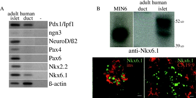
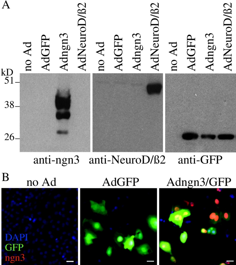
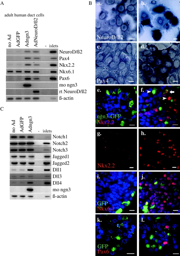
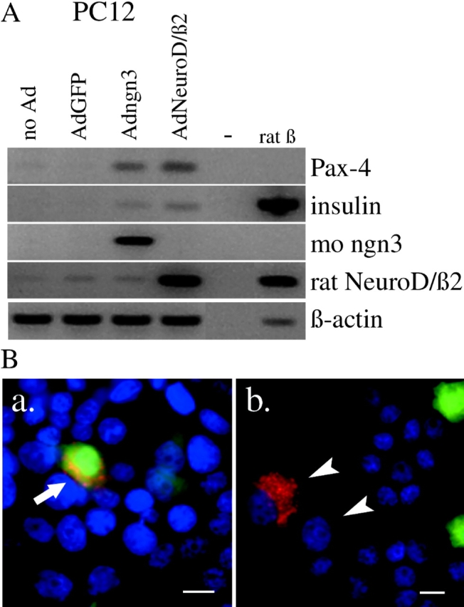
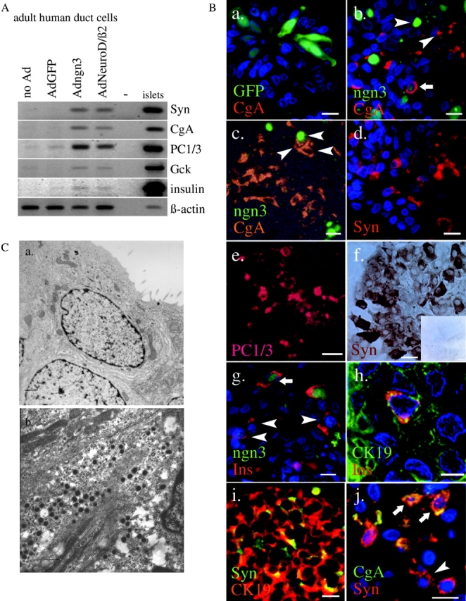
Regulatory proteins have been identified in embryonic development of the endocrine pancreas. It is unknown whether these factors can also play a role in the formation of pancreatic endocrine cells from postnatal nonendocrine cells. The present study demonstrates that adult human pancreatic duct cells can be converted into insulin-expressing cells after ectopic, adenovirus-mediated expression of the class B basic helix-loop-helix factor neurogenin 3 (ngn3), which is a critical factor in embryogenesis of the mouse endocrine pancreas. Infection with adenovirus ngn3 (Adngn3) induced gene and/or protein expression of NeuroD/beta2, Pax4, Nkx2.2, Pax6, and Nkx6.1, all known to be essential for beta-cell differentiation in mouse embryos. Expression of ngn3 in adult human duct cells induced Notch ligands Dll1 and Dll4 and neuroendocrine- and beta-cell-specific markers: it increased the percentage of synaptophysin- and insulin-positive cells 15-fold in ngn3-infected versus control cells. Infection with NeuroD/beta2 (a downstream target of ngn3) induced similar effects. These data indicate that the Delta-Notch pathway, which controls embryonic development of the mouse endocrine pancreas, can also operate in adult human duct cells driving them to a neuroendocrine phenotype with the formation of insulin-expressing cells.


Figures

Figure 1.
Figure 1.
Endogenous expression of key developmental transcription factors in adult human pancreatic duct cells. (A) RT-PCR analysis of RNA extracted from adult human duct cells compared with adult human islet cells. (B) Nkx6.1 protein in adult human pancreas as determined by immunoblot of extracts from enriched duct or islet cells (MIN6 were control cells), and immunostaining on sections of human donor pancreas. Bars, 10 μm.
Figure 2.
Figure 2.
Specificity and efficiency of adenovirus-mediated transgene expression. Immunoblotted protein extracts (2 μg total protein) (A) and immunostained monolayers (B) of noninfected and virally infected adult human pancreatic duct cells. Bars, 10 μm.
Figure 3.
Figure 3.
Effect of adenovirus-mediated ectopic expression of ngn3 or NeuroD/β2 on key transcription factors and signal transduction proteins in adult human duct cells. (A) RT-PCR analysis of RNA encoding key developmental transcription factors from control and virus-infected duct cells and islets. Adngn3-expressed mouse ngn3 and AdNeuroD/β2-expressed rat NeuroD/β2. (B) Analysis of the effects of ngn3 (b, d, f, h, j, and l) compared with AdGFP-infected control cells (a, c, e, g, i, and k) at the cellular level by in situ hybridization of NeuroD/β2 (a and b) and Pax4 (c and d) mRNA and immunofluorescence for Nkx2.2 (e–h), ngn3 (e and f), Nkx6.1 (i and j) and Pax6 (k and l). Sections for immunocytochemistry underwent short (1 h) fixation to preserve GFP fluorescence. Arrowheads in panel f point to cells expressing either Nkx2.2 or ngn3 (intense red, respectively, green nuclear staining), and arrow points to a cell expressing Nkx2.2 and still containing GFP (weak green fluorescence in cytoplasm combined with weak red staining in the nucleus) without high level expression of ngn3. Panels g and h represent a combination of phase–contrast and fluorescence microscopy (Nkx2.2 immunostaining), emphasizing the massive effect of ngn3. Bars, 10 μm. (C) RT-PCR analysis of RNA encoding Notch, Jagged, and Delta isoforms from control and virus-infected duct cells and human islets.
Figure 4.
Figure 4.
Effect of adenovirus-mediated expression of ngn3 or NeuroD/β2 in the neuroendocrine cell line PC12. (A) RT-PCR analysis of RNA encoding Pax4 and insulin from control and virus-infected PC12 cells and purified rat β-cells. (B) Immunostaining of insulin in PC12 cells that were infected at low MOI with Adngn3. Arrow points to insulin-positive PC12 cell that still contains lots of active GFP; arrowheads point to PC12 cells that express insulin but contain no active GFP or ngn3 anymore. Bars, 10 μm.
Figure 5.
Figure 5.
Effect of adenovirus-mediated ectopic expression of ngn3 or NeuroD/β2 on markers of the differentiated endocrine phenotype in adult human duct cells. (A) RT-PCR analysis of RNA encoding endocrine marker proteins in adult human duct cells and islet cells. (B) Immunostaining of control (AdGFP-) (a and f, inset) and Adngn3- (b and j) infected duct cells. Nuclei stained blue by DAPI (a, b, d, g, h, and j). All immunoreactions were labeled with a fluorescent secondary antibody except for panel f which was immunochemically stained (ABC peroxidase). Panel j represents a double labeling for CgA (FITC) and Syn (Cy3). Panels c and e represent a combination of phase– contrast and fluorescence microscopy. Noninfected and control-infected duct cells contained a low number of endocrine cells (<1% insulin positivity and <2% synaptophysin positivity). Endocrine marker proteins were chromogranin A (CgA), synaptophysin (Syn), prohormone convertase 1/3 (PC1/3), and insulin (Ins); duct cell marker was CK19. Noninfected and GFP control- infected duct cells contained none of the endocrine proteins under study as determined by costaining for the duct cell markers CK19 or CA19.9. Bars, 10 μm. Sections for immunocytochemistry underwent overnight fixation which abolished GFP fluorescence. Arrowheads, single positive cells on panels b and c (either ngn3 or CgA), g (insulin), and j (Syn); arrows, cells coexpressing CgA and ngn3 (b), insulin and ngn3 (g), or CgA and Syn (j). (C) Electron micrograph of control (a) and transdifferentiating (b) adult human pancreatic duct cells 10 d postinfection with AdGFP (a) or Adngn3 (b). Adngn3-infected cells display 180-nm secretory granules with a homogenous, electron dense matrix. Bars, 1 μm.

References

    1. Apelqvist, A., H. Li, L. Sommer, P. Beatus, D.J. Anderson, T. Honjo, M. Hrabe de Angelis, U. Lendahl, and H. Edlund. 1999. Notch signalling controls pancreatic cell differentiation. Nature. 400:877–881. - PubMed
    1. Artavanis-Tsakonas, S., M.D. Rand, and R.J. Lake. 1999. Notch signaling: cell fate control and signal integration in development. Science. 284:770–776. - PubMed
    1. Assady, S., G. Maor, M. Amit, J. Itskovitz-Eldor, K.L. Skorecki, and M. Tzukerman. 2001. Insulin production by human embryonic stem cells. Diabetes. 50:1691–1697. - PubMed
    1. Atouf, F., P. Czernichow, and R. Scharfmann. 1997. Expression of neuronal traits in pancreatic beta cells. Implication of neuron-restrictive silencing factor/repressor element silencing transcription factor, a neuron-restrictive silencer. J. Biol. Chem. 272:1929–1934. - PubMed
    1. Bonner-Weir, S., M. Taneja, G.C. Weir, K. Tatarkiewicz, K.H. Song, A. Sharma, and J.J. O'Neil. 2000. In vitro cultivation of human islets from expanded ductal tissue. Proc. Natl. Acad. Sci. USA. 97:7999–8004. - PMC - PubMed

Publication types

MeSH terms