. 2002 Oct 28;159(2):313-24.
doi: 10.1083/jcb.200203117. Epub 2002 Oct 28.

Down-regulation of Delta by proteolytic processing

Affiliations

Down-regulation of Delta by proteolytic processing

Ketu Mishra-Gorur et al. J Cell Biol. .

Abstract

Notch signaling regulates cell fate decisions during development through local cell interactions. Signaling is triggered by the interaction of the Notch receptor with its transmembrane ligands expressed on adjacent cells. Recent studies suggest that Delta is cleaved to release an extracellular fragment, DlEC, by a mechanism that involves the activity of the metalloprotease Kuzbanian; however, the functional significance of that cleavage remains controversial. Using independent functional assays in vitro and in vivo, we examined the biological activity of purified soluble Delta forms and conclude that Delta cleavage is an important down-regulating event in Notch signaling. The data support a model whereby Delta inactivation is essential for providing the critical ligand/receptor expression differential between neighboring cells in order to distinguish the signaling versus the receiving partner.


Figures
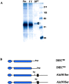
Figure 1.
Figure 1.
Purification of DlEC and schematic of Delta constructs. (A) DlEC was purified from conditioned medium from S2 cells stably expressing Delta using a Delta antibody (9B) affinity column. The purified form runs as a doublet as seen by SDS-PAGE analysis and silver staining. (B) Schematic representation of Delta constructs used to analyze the function of DlEC.
Figure 2.
Figure 2.
Expression of Delta mutant constructs. (A) Both the DlEC581 and DlEC593 truncated mutant constructs elicit a soluble protein into the medium. DlEC593 is slightly larger than DlEC581 consistent with the predicted 11 amino acid difference in their sequence. (B) The mutant constructs of Delta transfected into S2 cells and analyzed for Delta cleavage show that Serine mutants of Delta undergo cleavage like WT Delta. Full-length Delta (DlFL) was detected in the cell lysates (C), whereas DlEC is detected in the medium (M) by Western analysis with 9B antibody.
Figure 3.
Figure 3.
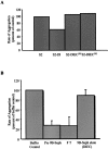
DlEC does not interact with Notch in S2-N cells. (A) Preincubation of S2-N cells with concentrated conditioned medium from S2 cells stably expressing DlEC581 or DlEC593 does not affect the rate of aggregation of S2-N and S2-Dl cells. Preincubation with concentrated conditioned medium from S2-Dl cells significantly affects the rate of aggregation of S2-N and S2-Dl cells. (B) The pre, flow-through (FT) and DlEC fractions (of 9B-affinity purification, Fig. 1 A) were analyzed in the N-Dl cell aggregation as described in the methods. Essentially all of the inhibitory activity of the precolumn sample is seen in the F.T. DlEC shows no significant inhibition of aggregation at the highest concentrations examined (up to 0.58 μM).
Figure 4.
Figure 4.
Fractionation of N-Dl cell aggregation inhibitory activity. (A) A sample of concentrated S2-Dl cell condition medium was fractionated on a Superdex 200HR size exclusion column. (B) Fractions analyzed by Western blotting with the 9B antibody reveal the DlEC protein in the precolumn sample and eluted fractions with a peak at ∼12.7 ml. A faint band consistent in size with full-length Dl is seen in the precolumn sample (*) and elutes in the void volume (V0). The V0 fraction contains proteins in the size range of 600 kD. (C) Pooled fractions were analyzed for inhibition of aggregation in the N-Dl cell aggregation assay. The precolumn sample shows significant inhibition of aggregation. Inhibition is still seen when the precolumn sample is diluted 1:10. Only pool 1, containing the V0 peak and the full-length Dl, showed inhibition of aggregation. This inhibition is equivalent to the 1:10 diluted precolumn sample, which is consistent with the approximate 10-fold dilution that the sample experiences during the run of the column. Fractions containing DlEC elicit no activity in the aggregation assay.
Figure 5.
Figure 5.
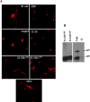
DlEC does not induce neurite retraction in cultured cortical neurons. (A) The effect of DlEC on primary cultured cortical neurons is shown in the representative images. There was no effect seen with 5× concentrated conditioned medium from S2 cells or PBS alone. Extensive neurite retraction associated with Notch activation is seen when the neurons are cocultured with cells expressing the Jagged ligand or treated with 5× concentrated conditioned medium from S2-Dl cells. No effect on neurite length is seen when neurons are treated with 5× concentrated conditioned medium from S2-DlEC581 or S2-DlEC593 or affinity purified DlEC. (B) Western analysis with anti-Delta 9B antibody on the conditioned medium from S2-Dl cells shows the presence of full length WT Delta in the medium.
Figure 6.
Figure 6.
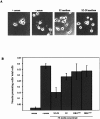
DlEC does not inhibit neurite outgrowth in neuroblastoma N2A cells. (A) Withdrawal of serum from actively growing N2A cells results in neurite outgrowth. This response is associated with Notch activation and shown in representative cultures in A. Addition of 5× concentrated conditioned medium from S2-Dl cells at the time of serum withdrawal inhibits neurite outgrowth. (B) The number of neurite presenting cells relative to the total number of cells in the plate is represented in the graph. Conditioned medium from S2, S2-DlEC581 and DlEC593 had a very mild effect on neurite outgrowth as compared with the effect of conditioned medium from S2-Dl cells.
Figure 7.
Figure 7.
Full-length Delta, but not DlEC, induces E(Spl)m3 expression in S2-N cells. (A) S2-N cells were aggregated with live S2-Dl cells for various time periods as described in Materials and methods. The cells were harvested and RNA was used for RT-PCR for either mβ, mγ, or m3 expression. The rp49 gene was used as an internal control. Unlike mβ and mγ only m3 transcription was seen to be specifically up-regulated in S2-N cells after 30 min of exposure to Delta expressing cells. (B) S2-N cells were aggregated with live S2-Dl cells or formalin fixed S2-Dl cells. The S2-N cells were also treated with 5× concentrated conditioned medium from S2-Dl cells or cocultured with S2-Dl cells in a transwell system. m3 expression was induced only when S2-N cells were aggregated with either live or fixed S2-Dl cells. There was no induction of m3 in the transwell system. The m3 induction with S2-Dl conditioned medium is correlated to the presence of full-length Dl in the medium (C). No m3 induction is seen when S2-N cells are aggregated with either live or fixed S2 cells. (C) Western analysis with anti-Delta 9B antibody of the S2-Dl conditioned medium and the medium in the lower and upper chambers of the transwell system. DlEC is found in the lower chamber with the S2-N cells within 30 min of culturing.
Figure 7.
Figure 7.
Full-length Delta, but not DlEC, induces E(Spl)m3 expression in S2-N cells. (A) S2-N cells were aggregated with live S2-Dl cells for various time periods as described in Materials and methods. The cells were harvested and RNA was used for RT-PCR for either mβ, mγ, or m3 expression. The rp49 gene was used as an internal control. Unlike mβ and mγ only m3 transcription was seen to be specifically up-regulated in S2-N cells after 30 min of exposure to Delta expressing cells. (B) S2-N cells were aggregated with live S2-Dl cells or formalin fixed S2-Dl cells. The S2-N cells were also treated with 5× concentrated conditioned medium from S2-Dl cells or cocultured with S2-Dl cells in a transwell system. m3 expression was induced only when S2-N cells were aggregated with either live or fixed S2-Dl cells. There was no induction of m3 in the transwell system. The m3 induction with S2-Dl conditioned medium is correlated to the presence of full-length Dl in the medium (C). No m3 induction is seen when S2-N cells are aggregated with either live or fixed S2 cells. (C) Western analysis with anti-Delta 9B antibody of the S2-Dl conditioned medium and the medium in the lower and upper chambers of the transwell system. DlEC is found in the lower chamber with the S2-N cells within 30 min of culturing.
Figure 8.
Figure 8.
Mild eye and wing phenotypes associated with DlEC in transgenic flies. (top) Scanning electron micrograph (SEM) of adult eyes. Expression of DlEC581 and DlEC593 driven by the GMR promoter yields a phenotype similar to a WT eye with occasional miss positioning or duplication of bristles (arrows). Expression of Ala581Ser, Ala593Ser, and WT Delta with the same driver results in duplication of bristles and melting and smoothing of lens material. Tangential sections show extra photoreceptors (arrowheads) as well as extra interommatidial pigment cells (*) in the Ala581Ser, Ala593Ser, and WT Delta eyes. (bottom) Adult wing phenotypes resulting from expression of Delta mutants: DlEC581 and DlEC593 exhibited WT wings with the Vg-Gal4 driver, whereas mild wing deltas and occasional extra vein material was seen with the A9-Gal4 driver. Expression of WT Delta under either the Vg- or A9-driver resulted in rudimentary wings. The Ala581Ser or Ala593Ser mutants resulted in phenotypes identical to those seen with WT Delta (not depicted).
Figure 8.
Figure 8.
Mild eye and wing phenotypes associated with DlEC in transgenic flies. (top) Scanning electron micrograph (SEM) of adult eyes. Expression of DlEC581 and DlEC593 driven by the GMR promoter yields a phenotype similar to a WT eye with occasional miss positioning or duplication of bristles (arrows). Expression of Ala581Ser, Ala593Ser, and WT Delta with the same driver results in duplication of bristles and melting and smoothing of lens material. Tangential sections show extra photoreceptors (arrowheads) as well as extra interommatidial pigment cells (*) in the Ala581Ser, Ala593Ser, and WT Delta eyes. (bottom) Adult wing phenotypes resulting from expression of Delta mutants: DlEC581 and DlEC593 exhibited WT wings with the Vg-Gal4 driver, whereas mild wing deltas and occasional extra vein material was seen with the A9-Gal4 driver. Expression of WT Delta under either the Vg- or A9-driver resulted in rudimentary wings. The Ala581Ser or Ala593Ser mutants resulted in phenotypes identical to those seen with WT Delta (not depicted).
Figure 9.
Figure 9.
A functional model for Delta cleavage. Notch and Delta are initially expressed equivalently on the cell surface. Delta (Dl) is cleaved and consequently inactivated either directly or indirectly by Kuzbanian (Kuz). The ligand is down-regulated so as to no longer interact with the Notch receptor in the same or adjacent cells; Delta removal is reinforcing the “signal receiving status” of that cell. Thus, the Notch receptor (N) can interact with the ligands in the adjacent, “signaling” cell. Although the model implies that Kuz is differentially regulated between critical neighbors, this hypothesis is yet to be tested. Furthermore, the model allows for the possibility of a positive interaction between Kuz and Notch, which has been postulated by some studies.

Similar articles

Cited by

References

    1. Al-Atia, G.R., P. Fruscoloni, M. Jacobs-Lorena. 1985. Translational regulation of mRNAs for ribosomal proteins during early Drosophila development. Biochemistry. 24:5798–5803. - PubMed
    1. Artavanis-Tsakonas, S., K. Matsuno, and M.E. Fortini. 1995. Notch signaling. Science. 268:225–232. - PubMed
    1. Artavanis-Tsakonas, S., M.D. Rand, and R.J. Lake. 1999. Notch signaling: cell fate control and signal integration in development. Science. 284:770–776. - PubMed
    1. Bourouis, M., and B. Jarry. 1983. Vectors containing a prokaryotic dihydrofolate reductase gene transform Drosophila cells to methotrexate-resistance. EMBO J. 2:1099–1104. - PMC - PubMed
    1. Brand, A.H., and N. Perrimon. 1993. Targeted gene expression as a means of altering cell fates and generating dominant phenotypes. Development. 118:401–415. - PubMed

Publication types

MeSH terms