. 2002 Oct 28;159(2):361-72.
doi: 10.1083/jcb.200207050. Epub 2002 Oct 28.

Distinct claudins and associated PDZ proteins form different autotypic tight junctions in myelinating Schwann cells

Affiliations

Distinct claudins and associated PDZ proteins form different autotypic tight junctions in myelinating Schwann cells

Sebastian Poliak et al. J Cell Biol. .

Abstract

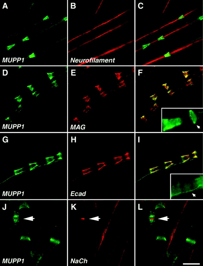
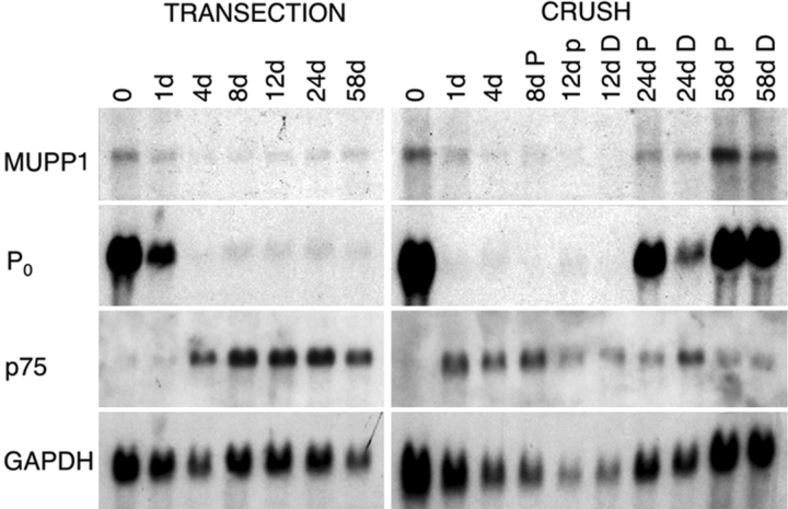
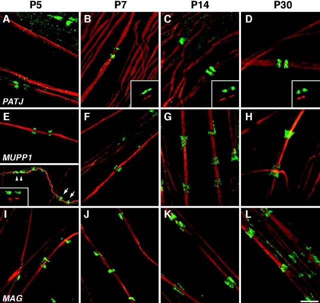
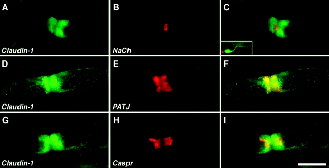
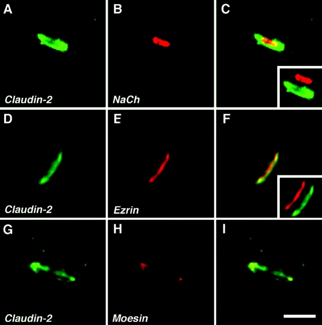
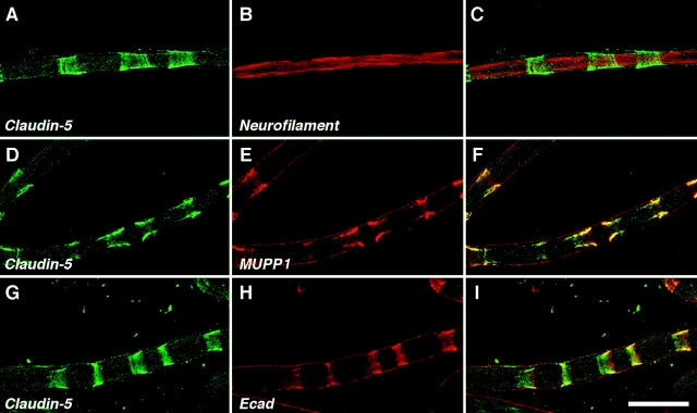
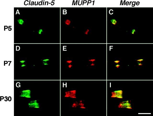
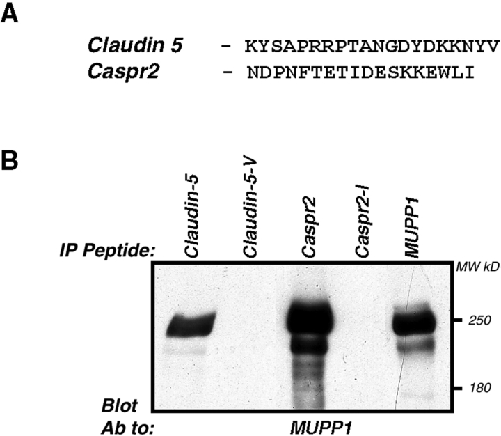
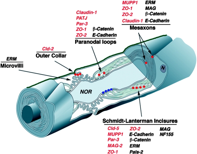
The apposed membranes of myelinating Schwann cells are joined by several types of junctional specializations known as autotypic or reflexive junctions. These include tight, gap, and adherens junctions, all of which are found in regions of noncompact myelin: the paranodal loops, incisures of Schmidt-Lanterman, and mesaxons. The molecular components of autotypic tight junctions have not been established. Here we report that two homologues of Discs Lost-multi PDZ domain protein (MUPP)1, and Pals-associated tight junction protein (PATJ), are differentially localized in myelinating Schwann cells and associated with different claudins. PATJ is mainly found at the paranodal loops, where it colocalized with claudin-1. MUPP1 and claudin-5 colocalized in the incisures, and the COOH-terminal region of claudin-5 interacts with MUPP1 in a PSD-95/Disc Large/zona occludens (ZO)-1 (PDZ)-dependent manner. In developing nerves, claudin-5 and MUPP1 appear together in incisures during the first postnatal week, suggesting that they coassemble during myelination. Finally, we show that the incisures also contain four other PDZ proteins that are found in epithelial tight junctions, including three membrane-associated guanylate-kinase proteins (membrane-associated guanylate-kinase inverted-2, ZO-1, and ZO-2) and the adaptor protein Par-3. The presence of these different tight junction proteins in regions of noncompact myelin may be required to maintain the intricate cytoarchitecture of myelinating Schwann cells.


Figures

Figure 1.
Figure 1.
Domain organization and subcellular localization of MUPP1 and PATJ in epithelial cells. (A) Schematic structure of Drosophila DLT and its mammalian homologues, MUPP1 and PATJ. All three proteins are composed of several PDZ domains (numbered squares), as well as a Lin7 binding domain (ellipses) in their NH2 terminus. The regions used to generate different antibodies against PATJ and MUPP1 are indicated, along with the name of each antibody. (B) Immunoprecipitation of MUPP1 and PATJ. HEK-293 cells transfected with MUPP1 (left) or PATJ (right) were incubated with preimmune serum (CS) or the indicated antibodies (IP Ab:). Precipitated proteins were immunoblotted using an antibody to MUPP1 or PATJ as indicated. The multiple bands detected in the left panel are degradation products of MUPP1. (C) PATJ and MUPP1 are localized at tight junctions in MDCK cells. Double immunofluorescence staining of MDCK cells using rabbit antisera (green) against PATJ or MUPP1 and monoclonal antibodies (red) against E-cadherin (Ecad), or using mouse antisera (green) against PATJ or MUPP1 and rabbit antisera (red) against ZO-1 or claudin-1 as indicated. The merged images are shown in the right panels (C, F, I, and L); insets show higher magnification of the small square labeled in each panel. Note that MUPP1 and PATJ colocalize with claudin-1 and ZO-1 but not E-cadherin. Bar, 10 μm.
Figure 2.
Figure 2.
MUPP1 is located at Schmidt-Lanterman incisures. (A–I) Images of teased fibers from adult rat sciatic nerves, double labeled with an antiserum against MUPP1 (green) and a monoclonal antibody (red) against neurofilament, MAG, or E-cadherin (Ecad) as indicated. For each labeling, the right panel shows the merge image. The insets in F and I show the expression of MUPP1 in annular ribbons and the mesaxon, respectively. (J–L) Images of teased fibers from mouse sciatic nerve labeled with an antiserum against MUPP1 (green) and a monoclonal antibody (red) to voltage-gated Na+ channels (NaCh). MUPP1 was detected in incisures and paranodes; the nodal Na+ channels are indicated (arrow). The merged image is shown in L. Bars: A–I, 20 μm; J–L, 15 μm.
Figure 3.
Figure 3.
Northern blot analysis of MUPP1 expression after transection and crush injury. Each lane contains an equal amount (10 μg) of total RNA isolated from the distal stumps of sciatic nerves that had been transected or crushed. The number of days after each of these lesions are indicated; the 0 time point is from unlesioned adult nerves. In crushed nerves, the distal nerve-stumps were divided into proximal (P) and distal (D) segments of equal lengths. The blot was sequentially probed with radiolabeled cDNA for MUPP1, p75, GAPDH, and P0, and exposed to film for 14 d, 14 d, 4 d, and 4 h, respectively.
Figure 4.
Figure 4.
PATJ is mainly found in the paranodal loops and only weakly in Schmidt-Lanterman incisures. Images of teased fibers from adult rat sciatic nerves, double labeled with an antiserum to PATJ (green) and a monoclonal antibody (red) to neurofilament, Caspr, or E-cadherin (Ecad). The merged images are shown on the right panel of each row. PATJ labeling surrounded that of Caspr, which marks the paranodal membrane of axons. PATJ antibody often labeled the incisures closest to the node (G and I, arrows), but was missing from all other E-cadherin–labeled incisures (I). Some weak staining of incisures was occasionally detected (J and L, arrows). Bar, 10 μm.
Figure 5.
Figure 5.
Developmental analysis of MUPP1 and PATJ in myelinated peripheral nerve. Images of teased fibers from rat sciatic nerves at the indicated ages, double labeled with a monoclonal antibody to neurofilaments (red) and an antiserum to PATJ, MUPP1, or MAG (green). PATJ was present in the paranodal loops at all ages examined. At P5 and P7, strong MUPP1 staining (green) was detected in the paranodal region (arrowheads in lower half of E) and incisures (arrows). Note that during the first postnatal week, the incisures were not fully developed and appear as narrow rings. As development progressed, MUPP1 staining remains strong in incisures but not in paranodes. The insets in B-E show shifted images to illustrate that PATJ or MUPP1 staining (green) surrounds Caspr staining (red). Bar, 10 μm.
Figure 6.
Figure 6.
Claudin-1 is located at the paranodal loops and along the mesaxon. Double immunofluorescence labeling of teased mouse sciatic nerve fibers using antibodies to claudin-1 (green) and Na+ channels (A–C, NaCh), PATJ (D–F), or Caspr (G–I), all in red. The right panel of each row shows the merged image. Claudin-1 was found in the paranodal region at both sides of the node (A–C), and colocalized with PATJ (D–F). Note that claudin-1 and Caspr are localized in distinct parts of the paranode (G–I). The inset in panel C shows internodal staining of claudin-1; this was more pronounced in smaller axons. Bar, 15 μm.
Figure 7.
Figure 7.
Localization of claudin-2 to the nodal region in peripheral nerves. Images of teased fibers from adult mouse sciatic nerve, double labeled for claudin-2 (green) and voltage-gated Na+ channels (A–C, NaCh), ezrin (D–F), or moesin (G–I), all in red. The left panel of each row shows the merged images. The insets in C and F show merged images in which the red and the green channels were shifted apart. Note that nodal membranes were labeled for NaCh, and were surrounded by a ring of claudin-2 staining. Claudin-2 colocalized with ezrin and moesin, which labeled Schwann cell microvilli. Bar, 5 μm.
Figure 8.
Figure 8.
Colocalization of claudin-5 and MUPP1 in incisures. Images of teased fibers from adult mouse sciatic nerves, double labeled for claudin-5 (green) and neurofilaments, MUPP1 or E-cadherin (Ecad), all in red. The right panel shows the merged images. Note that claudin-5 and MUPP1 are highly colocalized in incisures. The coating of MUPP1 and E-cadherin on the outside of the teased fibers is nonspecific, resulting from staining mouse teased fibers with a mouse monoclonal antibody. Bar, 25 μm.
Figure 9.
Figure 9.
MUPP1 and claudin-5 appear together in developing myelinated nerves. These are images of teased fibers from rat sciatic nerves at the indicated ages, double labeled for MUPP1 (green) and claudin-5 (red). Note that claudin-5 and MUPP1 colocalized in incisures at all ages. Bar, 10 μm.
Figure 10.
Figure 10.
MUPP1 bind to the carboxyl terminus of claudin-5. (A) Amino acid sequences of the COOH-termini of claudin-5 and Caspr2 used in the binding experiments. Note that both proteins contain a sequence that may bind type-II PDZ domains. (B) The immobilized peptides shown in A, or the same peptides lacking the last amino acid, were mixed with lysates of HEK-293 cells expressing MUPP1. Purified complexes were separated on SDS-gel and immunoblotted using an antibody to MUPP1. Immunoprecipitation with MUPP1 antibody (MUPP1 IP) was used as a positive control. The location of molecular mass markers in kD is shown on the right.
Figure 11.
Figure 11.
Location of autotypic junction proteins in myelinating Schwann cells. The organization of peripheral myelinated nerve is shown schematically. The location of autotypic junctions in the Schmidt Lanterman incisures, paranodal loops, mesaxons and the outer aspect of the nodal gap are marked with red dots. The heterotypic septate-like junction formed between the axon and the paranodal loops of the myelinating Schwann cell is labeled with blue dots. The basal lamina covering the Schwann cell-axon unit is shown in green. The localization of different proteins discussed in this study is listed. Tight junction proteins are labeled in red. Note that autotypic tight junctions present in different aspects of noncompact myelin contain distinct junctional complexes (modified from Spiegel and Peles [2002] and used with permission of Taylor and Francis, www.tandf.co.uk).

Similar articles

Cited by

References

    1. Altevogt, B.M., K.A. Kleopa, F.R. Postma, S.S. Scherer, and D.L. Paul. 2002. Cx29 is uniquely distributed within myelinating glial cells of the central and peripheral nervous systems. J. Neurosci. 15:6458–6470. - PMC - PubMed
    1. Balice-Gordon, R.J., L.J. Bone, and S.S. Scherer. 1998. Functional gap junctions in the Schwann cell myelin sheath. J. Cell Biol. 142:1095–1104. - PMC - PubMed
    1. Barritt, D.S., M.T. Pearn, A.H. Zisch, S.S. Lee, R.T. Javier, E.B. Pasquale, and W.B. Stallcup. 2000. The multi-PDZ domain protein MUPP1 is a cytoplasmic ligand for the membrane-spanning proteoglycan NG2. J. Cell. Biochem. 79:213–224. - PMC - PubMed
    1. Baumgartner, S., J.T. Littleton, K. Broadie, M.A. Bhat, R. Harbecke, J.A. Lengyel, R. Chiquet-Ehrismann, A. Prokop, and H.J. Bellen. 1996. A Drosophila neurexin is required for septate junction and blood-nerve barrier formation and function. Cell. 87:1059–1068. - PubMed
    1. Bazzoni, G., O.M. Martinez-Estrada, F. Orsenigo, M. Cordenonsi, S. Citi, and E. Dejana. 2000. Interaction of junctional adhesion molecule with the tight junction components ZO-1, cingulin, and occludin. J. Biol. Chem. 275:20520–20526. - PubMed

Publication types

MeSH terms