. 2002 Nov 12;99(23):14994-9.
doi: 10.1073/pnas.222425399. Epub 2002 Oct 25.

Mutations in Mcoln3 associated with deafness and pigmentation defects in varitint-waddler (Va) mice

Affiliations

Mutations in Mcoln3 associated with deafness and pigmentation defects in varitint-waddler (Va) mice

Federica Di Palma et al. Proc Natl Acad Sci U S A. .

Abstract

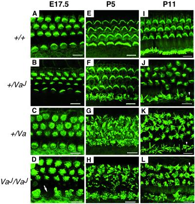
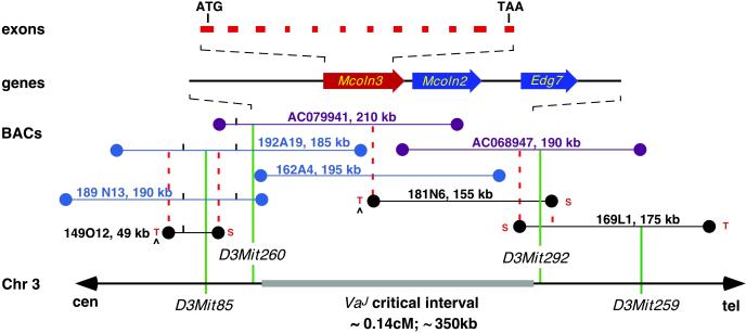
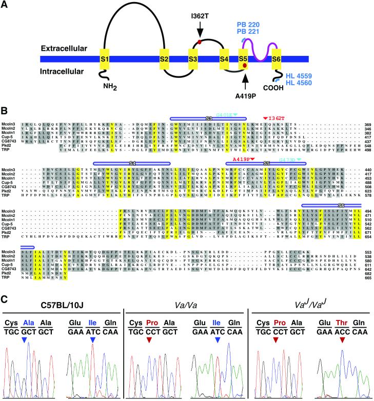
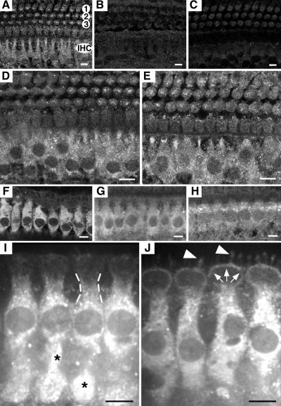
Deafness in spontaneously occurring mouse mutants is often associated with defects in cochlea sensory hair cells, opening an avenue to systematically identify genes critical for hair cell structure and function. The classical semidominant mouse mutant varitint-waddler (Va) exhibits early-onset hearing loss, vestibular defects, pigmentation abnormalities, and perinatal lethality. A second allele, Va(J), which arose in a cross segregating for Va, shows a less severe phenotype. By using a positional cloning strategy, we identify two additional members of the mucolipin gene family (Mcoln2 and Mcoln3) in the 350-kb Va(J) minimal interval and provide evidence for Mcoln3 as the gene mutated in varitint-waddler. Mcoln3 encodes a putative six-transmembrane-domain protein with sequence and motif similarities to the family of nonselective transient-receptor-potential (TRP) ion channels. In the Va allele an Ala419Pro substitution occurs in the fifth transmembrane domain of Mcoln3, and in Va(J), a second sequence alteration (Ile362Thr) occurring in cis partially rescues the Va allele. Mcoln3 localizes to cytoplasmic compartments of hair cells and plasma membrane of stereocilia. Hair cell defects are apparent by embryonic day 17.5, assigning Mcoln3 an essential role during early hair cell maturation. Our data suggest that Mcoln3 is involved in ion homeostasis and acts cell-autonomously. Hence, we identify a molecular link between hair cell physiology and melanocyte function. Last, MCOLN2 and MCOLN3 are candidate genes for hereditary and/or sporadic forms of neurosensory disorders in humans.


Figures

Fig 1.
Fig 1.
Development of cochlear hair cells in varitint-waddler mutants. Labeling of cochlear hair cells F-actin by FITC-phalloidin at E17.5 (A–D), P5 (E–H), and P11 (I–L). Shown are optical sections through the OC at the level of the stereocilia. Normal hair cells can be distinguished by E17.5 as a regular array of microvilli from which ordered rows of stereocilia bundles develop; in wild-type mice, hair cell stereocilia are V-shaped in OHCs (top three rows), and straight in IHCs (bottom row) (A, E, and I). In +/VaJ mutants hair cell bundles appear mostly normal at E17.5 (B); disorganization of IHC stereocilia is severe by birth and continues to progress from P5 (F) to P11 (J); only minor signs of disorganization are noticeable in OHC bundles by P5 (F), but disorganization of the OHC bundle continues to progress, becoming more pronounced by P11 (J). In +/Va mutants, IHC and OHC stereociliar bundles are equally disorganized at all time points (C, G, and K). In VaJ/VaJ mutants, stereocilia disorganization is apparent in both IHCs and OHCs at E17.5 (D), and progresses from P5 (H) to P11 (L). In all mutant genotypes, by P11 many stereocilia appear missing and extensive fusion and clumping are apparent (JL, arrowheads). The spatial organization of hair cells in the OC in all mutants remains unaltered with three rows of OHCs and one row of IHCs. (Bars, 10 μm.)
Fig 2.
Fig 2.
Physical map of the VaJ critical region. The thick horizontal gray line (Bottom) represents the VaJ critical interval defined by the polymorphic Mit markers on mouse chromosome 3. cM, centimorgan. A contig of 15 overlapping BAC clones is represented by horizontal lines; RPCI-23 BAC clones are shown in blue, Incyte Genomics BAC clones are in black, and BAC clones used for sample sequencing are in purple. Coordinates and corresponding sizes appear next to each BAC clone. SP6 and T7 ends of BACs are indicated by S or T, respectively. BAC ends used to derive probes for screening of the RPCI-23 BAC library are indicated by ^. Dashed lines in red indicate anchored SP6 and T7 BAC ends that have been used in PCR cross-screening and/or Southern blot experiments to confirm overlaps among BAC clones. Genes that map within the candidate region are shown with an arrow indicating transcription orientation. Mcoln3 exons are indicated. Genomic clones containing Mcoln2 and Mcoln3, (RP23-108E10), and (RP23-121J1).
Fig 3.
Fig 3.
Sequence analysis of Mcoln3. (A) Schematic representation of predicted structure of Mcoln3. Six putative transmembrane domains (S1–S6) and a putative pore region (amino acids 480–505; in purple) are shown. Mutations in Mcoln3 are indicated by red dots. Blue lines represent regions of Mcoln3 containing polyclonal antibody recognition sites: PB221 and PB220 antisera were raised against amino acids 446–462, HL4559, and HL4560 against amino acids 529–548. (B) clustalw alignment of the mucolipin family. Multiple sequence alignment of Mcoln3, Mcoln1, Mcoln2, C. elegans CUP-5, and Drosophila CG743 is shown with Pkd2 (amino acids 478–682) and TRP (amino acids 430–665). Putative transmembrane domains (S1–S6) are indicated by thick blue lines. Predicted ion transport domain and TRPL motif (PS50272) of Mcoln3 are located between amino acids 337–501 and amino acids 317–505, respectively. Conserved amino acids across the Mcoln3 TRPL region are shaded yellow. Sites of mutations in Mcoln3 are shown in red and in CUP-5 in teal. Amino acid positions are given on the right. (C) Mutation analysis. Sequence chromatographs showing nucleotide sequence and translation across the sites of mutations in C57BL/10J, Va/Va, and VaJ/VaJ genomic DNA. Nucleotide changes are shown in red.
Fig 4.
Fig 4.
Localization of Mcoln3 in mouse and rat OC by immunofluorescence. Shown are cross-sections through the OC showing the labeling of IHCs for Mcoln3. (A–E) Mouse OC at P11, and optical sections at the level of the cell body of IHCs. In A a single row of IHCs and three rows of OHCs (1, 2, and 3) are presented. (A, D, and E) PB221 labeling for Mcoln3. The cytoplasm of IHCs is intensively labeled in wild type (A), +/VaJ (D), and VaJ/VaJ (E); staining is absent after incubation with preimmune serum (B) and excess specific peptide (C). Some labeling is also observed in the cytoplasm of OHCs (A, D, and E), and under the cuticular plate (not shown). (FJ) Rat OC at 4 weeks of age, and optical sections at the level of cell bodies (FH) and the cuticular plate (I and J) of IHCs. All four anti-Mcoln3 antisera, PB220 (F and I), PB221 (J), HL4459 (G), and HL4460 (H) showed immunoreactivity in the cytoplasm of IHCs. In the cytoplasm, labeling appears as a punctuate pattern (*, I) throughout the IHC bodies, and it is also detected in the subcuticular region (dashed lines, I), the pericuticular necklace (white arrows, J), and in the IHC bundles (white arrowheads, J). Specificity of all four antisera was confirmed by immunoblot after bacterial expression of a partial Mcoln3 fusion protein (data not shown). Stereocilia staining was also observed on mouse OC (data not shown). (Bars, 80 μm.)

Comment in

  • Varitint-waddler: a double whammy for hearing.
    Steel KP. Steel KP. Proc Natl Acad Sci U S A. 2002 Nov 12;99(23):14613-5. doi: 10.1073/pnas.232585699. Epub 2002 Nov 4. Proc Natl Acad Sci U S A. 2002. PMID: 12417743 Free PMC article. No abstract available.

References

    1. Steel K. P. & Kros, C. J. (2001) Nat. Genet. 27 143-149. - PubMed
    1. Geissler E. N., Ryan, M. A. & Housman, D. E. (1988) Cell 55 185-192. - PubMed
    1. Chabot B., Stephenson, D. A., Chapman, V. M., Besmer, P. & Bernstein, A. (1988) Nature 335 88-89. - PubMed
    1. Matsui Y., Zsebo, K. M. & Hogan, B. L. (1990) Nature 347 667-669. - PubMed
    1. Huang E., Nocka, K., Beier, D. R., Chu, T. Y., Buck, J., Lahm, H. W., Wellner, D., Leder, P. & Besmer, P. (1990) Cell 63 225-233. - PubMed

Publication types

MeSH terms