. 2002 Nov;68(11):5241-8.
doi: 10.1128/AEM.68.11.5241-5248.2002.

Utilization of ganglioside-degrading Paenibacillus sp. strain TS12 for production of glucosylceramide

Affiliations

Utilization of ganglioside-degrading Paenibacillus sp. strain TS12 for production of glucosylceramide

Tomomi Sumida et al. Appl Environ Microbiol. 2002 Nov.

Abstract

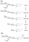
Gangliosides, sialic acid-containing glycosphingolipids, are membrane constituents of vertebrates and are known to have important roles in cellular differentiation, adhesion, and recognition. We report here the isolation of a bacterium capable of degrading gangliotetraose-series gangliosides and a new method for the production of glucosylceramide with this bacterium. GM1a ganglioside was found to be sequentially degraded by Paenibacillus sp. strain TS12, which was isolated from soil, as follows: GM1a --> asialo GM1 --> asialo GM2 --> lactosylceramide --> glucosylceramide. TS12 was found to produce a series of ganglioside-degrading enzymes, such as sialidases, beta-galactosidases, and beta-hexosaminidases. TS12 also produced beta-glucosidases, but glucosylceramide was somewhat resistant to the bacterial enzyme under the conditions used. Taking advantage of the specificity, we developed a new method for the production of glucosylceramide using TS12 as a biocatalyst. The method involves the conversion of crude bovine brain gangliosides to glucosylceramide by coculture with TS12 and purification of the product by chromatography with Wakogel C-300 HG.


Figures

FIG. 1.
FIG. 1.
Electron micrograph and phylogenetic tree of TS12. (A) TS12 cell stained with uranium acetate and observed with an electron microscope (JEM-1010). (B) Phylogenetic tree. Partial 16S rDNA fragments were amplified by PCR with universal primers. The nucleotide sequences were determined and then subjected to a phylogenetic analysis by using CLUSTAL W and 16S rDNA sequences of other Paenibacillus sp. obtained from a DNA database. The DDBJ accession numbers for the sequences used for phylogenetic comparisons are as follows: P. alvei, AJ320491; P. pabuli, X60630; P. amylolyticus, P85496; P. larvae subsp. pulvifaciens, AY030080, P. validus, AF353697; P. macerans, X57306; and P. polymyxa, AJ320493. The phylogenetic tree was constructed by the neighbor-joining method (24). The details are described in Materials and Methods.
FIG. 2.
FIG. 2.
Metabolic degradation of GM1a and NBD-GM1a during cultivation with TS12. TS12 cells were transferred into 250 μl of medium A and incubated at 30°C for different times. Twenty microliters of the culture supernatant was withdrawn every 6 h and analyzed by TLC with solvent I. (A) TLC stained with orcinol-H2SO4 reagent. AsGM1, asialo GM1; TDC, taurodeoxycholate. (B) Quantification of the compounds shown in panel A with a Shimadzu CS-9300PC chromatoscanner with the reflectance mode set at 540 nm. The values are means ± standard deviations for triplicate determinations. (C) Degradation of NBD-GM1a. One hundred picomoles of fluorescent GM1a was incubated with TS12 at 30°C for 3 days in medium A. The reaction product was separated by TLC by using solvent I. Lane 1, standard NBD-GM1a; lane 2, NBD-GM1a plus TS12; lane 3, standard NBD-GlcCer (NBD-C12:0); lane 4, standard NBD-Cer (NBD-C12:0).
FIG. 2.
FIG. 2.
Metabolic degradation of GM1a and NBD-GM1a during cultivation with TS12. TS12 cells were transferred into 250 μl of medium A and incubated at 30°C for different times. Twenty microliters of the culture supernatant was withdrawn every 6 h and analyzed by TLC with solvent I. (A) TLC stained with orcinol-H2SO4 reagent. AsGM1, asialo GM1; TDC, taurodeoxycholate. (B) Quantification of the compounds shown in panel A with a Shimadzu CS-9300PC chromatoscanner with the reflectance mode set at 540 nm. The values are means ± standard deviations for triplicate determinations. (C) Degradation of NBD-GM1a. One hundred picomoles of fluorescent GM1a was incubated with TS12 at 30°C for 3 days in medium A. The reaction product was separated by TLC by using solvent I. Lane 1, standard NBD-GM1a; lane 2, NBD-GM1a plus TS12; lane 3, standard NBD-GlcCer (NBD-C12:0); lane 4, standard NBD-Cer (NBD-C12:0).
FIG. 3.
FIG. 3.
Degradation of GM1a by TS12 (A) and fluorescence (NBD)-labeled GM1a (B).
FIG. 4.
FIG. 4.
Degradation of NBD-GM1a by cell-free supernatant of TS12. NBD-GM1a (100 pmol) was incubated at 37°C for different times with 20 μl of cell-free supernatant of TS12 or another strain. The reaction products were analyzed by TLC as described in Materials and Methods. (A) Incubation for 12 h. Lane 1, standard NBD-GM1a (NBD-C12:0); lane 2, NBD-GM1a with TS12 cell-free supernatant; lane 3, standard NBD-GlcCer (NBD-C12:0); lane 4, standard NBD-Cer (NBD-C12:0); lanes 5 to 10, NBD-GM1a with cell-free supernatants from Paenibacillus strains (lane 5, P. alvei; lane 6, P. amylolyticus; lane 7, P. validus; lane 8, P. pabuli; lane 9, P. macerans; lane 10, P. polymyxa). AsGM2, asialo GM2. (B) Incubation for 24 h. Lane 1, standard NBD-GM1a; lane 2, NBD-GM1a with TS12 cell-free supernatant; lane 3, standard NBD-GlcCer; lane 4, standard NBD-Cer.
FIG. 5.
FIG. 5.
Purification of GlcCer from culture supernatant of Paenibacillus sp. strain TS12. Crude bovine brain gangliosides (25 mg) were incubated at 30°C for 3 days with TS12 in 50 ml of medium B. GlcCer was isolated from the TS12 culture supernatant by using a Wakogel C-300 HG column. The elutes were checked by TLC as described in Materials and Methods. Std, standard containing LacCer (lower band) and GlcCer (upper band).
FIG. 6.
FIG. 6.
Purity of the prepared GlcCer. Aliquots of the purified GlcCer and standard GlcCer (10 μg each) were analyzed by TLC by using solvent II and were visualized with orcinol-H2SO4 (29) (A) or Coomassie brilliant blue R-250 (22) (B). Lane 1, purified GlcCer; lane 2, standard GlcCer from spleens of patients with Gaucher disease.
FIG. 7.
FIG. 7.
TOF-MS analysis of the purified GlcCer. The analysis was conducted with a Voyager Vestec mass spectrometer (PerSeptive Biosystem) in the positive-ion mode with 2,5-dihydroxybenzoic acid as the matrix.

References

    1. Arita, M., M. Iwamori, T. Higuchi, and Y. Nagai. 1983. Negative ion fast atom bombardment mass spectrometry of gangliosides and asialo gangliosides: a useful method for the structural elucidation of gangliosides and related neutral glycosphingolipids. J. Biochem. (Tokyo) 94:249-256. - PubMed
    1. Boldin, S. A., and A. H. Futerman. 2000. Up-regulation of glucosylceramide synthesis upon stimulation of axonal growth by basic fibroblast growth factor. J. Biol. Chem. 275:9905-9909. - PubMed
    1. Brosius, J., M. L. Palmer, P. J. Kennedy, and H. F. Noller. 1978. Complete nucleotide sequence of a 16S ribosomal RNA gene from Escherichia coli. Proc. Natl. Acad. Sci. USA 75:4801-4805. - PMC - PubMed
    1. Buchanan, R. E., and N. E. Gibbons (ed.). 1974. Bergey's manual of determinative bacteriology, 8th ed. The Williams & Wilkins Co., Baltimore, Md.
    1. Falk, P., L. C. Hoskins, and G. Larson. 1990. Bacteria of human intestinal microflora produce glycosidases specific for lacto-series glycosphingolipids. J. Biochem. (Tokyo) 108:466-474. - PubMed

Publication types

MeSH terms

LinkOut - more resources