. 2002 Nov 12;99(23):15194-9.
doi: 10.1073/pnas.232087499. Epub 2002 Oct 29.

RNA-binding protein Musashi family: roles for CNS stem cells and a subpopulation of ependymal cells revealed by targeted disruption and antisense ablation

Affiliations

RNA-binding protein Musashi family: roles for CNS stem cells and a subpopulation of ependymal cells revealed by targeted disruption and antisense ablation

Shin-ichi Sakakibara et al. Proc Natl Acad Sci U S A. .

Abstract

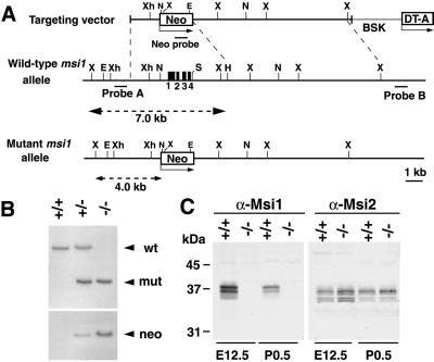
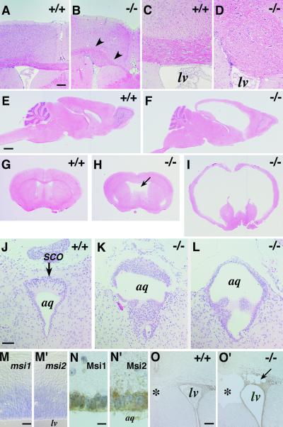
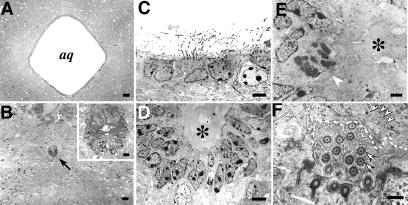
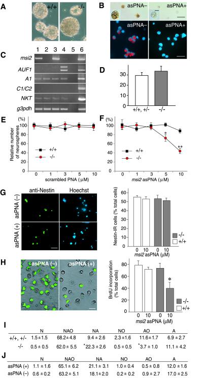
Homologues of the Musashi family of RNA-binding proteins are evolutionarily conserved across species. In mammals, two members of this family, Musashi1 (Msi1) and Musashi2 (Msi2), are strongly coexpressed in neural precursor cells, including CNS stem cells. To address the in vivo roles of msi in neural development, we generated mice with a targeted disruption of the gene encoding Msi1. Homozygous newborn mice frequently developed obstructive hydrocephalus with aberrant proliferation of ependymal cells in a restricted area surrounding the Sylvius aqueduct. These observations indicate a vital role for msi1 in the normal development of this subpopulation of ependymal cells, which has been speculated to be a source of postnatal CNS stem cells. On the other hand, histological examination and an in vitro neurosphere assay showed that neither the embryonic CNS development nor the self-renewal activity of CNS stem cells in embryonic forebrains appeared to be affected by the disruption of msi1, but the diversity of the cell types produced by the stem cells was moderately reduced by the msi1 deficiency. Therefore, we performed antisense ablation experiments to target both msi1 and msi2 in embryonic neural precursor cells. Administration of the antisense peptide-nucleotides, which were designed to specifically down-regulate msi2 expression, to msi1(-/-) CNS stem cell cultures drastically suppressed the formation of neurospheres in a dose-dependent manner. Antisense-treated msi1(-/-) CNS stem cells showed a reduced proliferative activity. These data suggest that msi1 and msi2 are cooperatively involved in the proliferation and maintenance of CNS stem cell populations.


Figures

Fig 1.
Fig 1.
Targeting strategy, germ-line transmission, and expression analysis of the msi1 gene. (A) Organization of the targeting vector, the msi1 gene, and the allele resulting from homologous recombination. Four exons (black boxes) of the msi1 allele containing the initiation codon were replaced with a Neo cassette. A 0.3-kb Sau3AI fragment (probe A) was used to screen for recombinant alleles, and the sizes of the recombinant and WT fragments after XbaI digestion are shown (broken lines). X, XbaI; E, EcoRV; Xh, XhoI; Not, NotI; S, SalI; H, HindIII; BSK, plasmid vector. (B) Germ-line transmission was confirmed by Southern blot analysis of XbaI- or EcoRV-digested tail DNA from a litter of F6 mice using probe A or the Neo probe, respectively. (C) Immunoblot analysis of brain lysates from E12.5 and P0.5 mice using anti-Msi1 or anti-Msi2 antibodies. Genotypes are indicated.
Fig 2.
Fig 2.
Development of hydrocephalus in postnatal msi1−/− mice with ependymal abnormalities and the concurrent expression of Msi1 and Msi2 in the developing CNS and aqueduct. Genotypes are indicated. (AI) Hematoxylin & eosin-stained adult brain. Agenesis of the CC observed in an adult msi1−/− mouse (B, arrowheads) compared with the normal CC in a WT littermate (A). (C and D) A higher magnification view of the normal CC and the Probst's bundle, respectively. (EI) Hydrocephalus of the adult msi1−/− mice. (E and F) Sagittal sections. (GI) Coronal sections at the level of the anterior commissure. (F and I) A severe hydrocephalic mutant showing massive ventricular dilation. (H) Moderate dilation of the lateral ventricles accompanied by cavitation of the septum pellucidum and hypoplasia of the septum. (JL) Coronal sections through the aqueduct of the P7 brain, showing abnormal accumulation and polyposis of ependymal cells surrounding the Sylvius aqueduct and SCO. (M and M′) mRNA in situ hybridization analysis of msi1 and msi2 in the WT E12.5 telencephalon showing their expression in the VZ. (N and N′) Immunohistochemical detection of Msi1 and Msi2 in ependymal cells surrounding the aqueduct of WT P3 mice. Immunoreactivities were visualized by DAB (brown), and the nuclei were counterstained with hematoxylin. (O and O′) Expression of Msi2 in the SVZ and ependymal cells lining the lateral ventricle of a hydrocephalic msi1−/− (O′) and a WT littermate (O) at P3. The msi1−/− tissue shows partial destruction of the SVZ (arrow). lv, lateral ventricle; aq, Sylvius aqueduct; *, septum pellucidum. (Scale bars: A and B, 150 μm; C, D, and JL, 50 μm; EI, 1 mm; M, 30 μm; N, 10 μm; O, 100 μm.)
Fig 3.
Fig 3.
Light microscopic and ultrastructural analyses of the msi1−/− ependyma at P14. (A and B) Light micrographs of the msi1−/− brain showing stenosis of the Sylvius aqueduct (B, arrow) and of the WT brain (A). Inset in B shows a low-power view of the ependymal cells in the aqueduct. (C) An electron micrograph of the WT aqueduct showing a single layer of ependymal cells and well organized microvilli protruding into the aqueductal lumen. (D) The aqueduct of an msi1−/− mouse, surrounded by the stratified ependymal epithelium composed of two or three cell layers. (E) A high-power view of msi1−/− ependymal cells, showing heterotopic mitotic figures without nuclear heteromorphism (arrowhead). Cytoplasmic organelles in these cells appear intact. The lumen of the msi1−/− aqueduct is completely filled with amorphous materials (asterisks in D and E). (F) A magnified view of msi1−/− ependymal evaginations and cilia. Cilia, characterized by central and peripheral tubules (arrows), are apposed to and are disarranged by solid materials (dashed line) that are abundant in the electron-dense particles of glycogen granules (arrowheads). (Scale bars: A and B, 40 μm; B Inset, 8 μm; C and D, 5 μm; E, 2 μm; F, 0.5 μm.)
Fig 4.
Fig 4.
Loss of msi1 and msi2 activities decreased the efficiency of neurosphere formation through the reduced proliferation of SFCs. (A) Neurospheres derived from the E14.5 telencephalons of msi1−/− or WT littermates (after three passages). (Scale bar, 100 μm.) (B) Suppression of Msi2 immunoreactivity on the asPNA-treated SFCs. Dissociated cells from the WT (Upper) or msi1−/− (Lower) primary neurospheres were incubated with or without asPNA (10 μM) for 24 h and immunostained with anti-Msi2 antibody. Immunoreactivities to Msi2 were visualized by a DAB reaction (brown, Upper) or Alaxa-568 (red, Lower). Insets are magnified views of representative cells, showing the repression of Msi2 protein expression without pyknotic changes. [Scale bar, 5 μm (Inset) or 25 μm (Upper and Lower).] (C) Semiquantitative RT-PCR analysis of msi2 mRNA, the msi-related genes encoding RNA-binding protein (AUF1, hnRNP A1, and hnRNP C1/C2), NKT mRNA, and an internal control mRNA (g3pdh). The msi1−/− or WT SFCs were incubated with or without asPNA for 24 h, then subjected to the RT-PCR. Lane 1, WT cells; lane 2, WT cells + asPNA; lane 3, msi1−/− cells; lane 4, msi1−/− cells + asPNA; lane 5, no RT control; lane 6, E14.5 WT cerebral cortex. (D) The number of neurospheres per field (×4) formed by the dissociated cells from msi1−/− (−/−, n = 3) or littermates (+/+, +/−, n = 3). (E and F) The number of neurospheres derived from the telencephalons of msi1−/− and their WT littermates in the presence of the scrambled PNA (E) or in the presence of the asPNA (F). The data were presented as the mean ± SEM (WT, n = ≈3–6; msi1−/−, n = ≈3–7). *P < 0.005, **P < 0.0001, in comparison with the WT control (unpaired t test). (G) The number of nestin+ cells after the exposure to asPNA for 24 h. Dissociated cells of the primary neurospheres derived from the WT and msi1−/− telencephalons were incubated with or without asPNA (10 μM) and then immunopositive cells for anti-nestin were counted (WT, n = 6; msi1−/−, n = 6). Photomicrographs showed the msi1−/− cells that were positive for nestin (green). (Scale bar, 25 μm.) (H) Decreased number of BrdU+ cells within the msi1−/− neurospheres exposed to asPNA. During the formation of neurospheres in the presence (+) or absence (−) of asPNA (10 μM), BrdU was administrated at 2 div. After the additional cultivation for 24 h, each sphere was dissociated and immunostained with anti-BrdU (WT, n = 6; msi1−/−, n = 6). Photomicrographs represented the BrdU-labeled msi1−/− cells (green). The data were presented as the mean ± SEM. *, P < 0.001 in comparison with the other conditions (two-tailed Student's t test). (Scale bar, 10 μm.) (I) Differentiation assay of neurospheres that derived from the msi1−/− (−/−) and their littermate cells (+/+, +/−). The differentiation capacity of each primary neurosphere was determined based on the cell types contained in each clone. N, neurons; A, astrocytes; O, oligodendrocytes. The majority (84.5%) of neurospheres from msi1−/− mice generated both neurons and glia (NAO+NA+NO). The clone types were analyzed for 98 spheres from WT or heterozygous mice (n = 5) and 234 spheres from msi1−/− mice (n = 10) and presented as the mean ± SEM. *, P < 0.0001, in comparison with WT and heterozygous controls (Student's t test). (J) Differentiation assay of neurospheres that derived from the msi1−/− cells treated with or without asPNA. The clone types were analyzed for 67 spheres from asPNA (+) and 141 spheres from asPNA (−).

Similar articles

Cited by

References

    1. Temple S. & Alvarez-Buylla, A. (1999) Curr. Opin. Neurobiol. 9, 135-141. - PubMed
    1. Sakakibara S., Imai, T., Hamaguchi, K., Okabe, M., Aruga, J., Nakajima, K., Yasutomi, D., Nagata, T., Kurihara, Y., Uesugi, S., et al. (1996) Dev. Biol. 176, 230-242. - PubMed
    1. Sakakibara S. & Okano, H. (1997) J. Neurosci. 17, 8300-8312. - PMC - PubMed
    1. Sakakibara S., Nakamura, Y., Satoh, H. & Okano, H. (2001) J. Neurosci. 21, 8091-8107. - PMC - PubMed
    1. Keyoung H. M., Roy, N. S., Benraiss, A., Louissaint, A., Jr., Suzuki, A., Hashimoto, K., Rashbaum, W. K., Okano, H. & Goldman, S. A. (2001) Nat. Biotechnol. 19, 843-850. - PubMed

Publication types

MeSH terms