. 2002 Nov 12;99(23):14946-51.
doi: 10.1073/pnas.232579599. Epub 2002 Oct 29.

The Usher syndrome proteins cadherin 23 and harmonin form a complex by means of PDZ-domain interactions

Affiliations

The Usher syndrome proteins cadherin 23 and harmonin form a complex by means of PDZ-domain interactions

Jan Siemens et al. Proc Natl Acad Sci U S A. .

Abstract

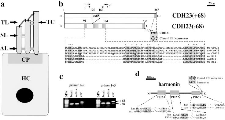
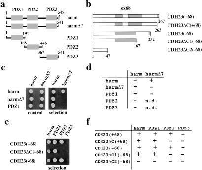
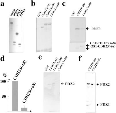
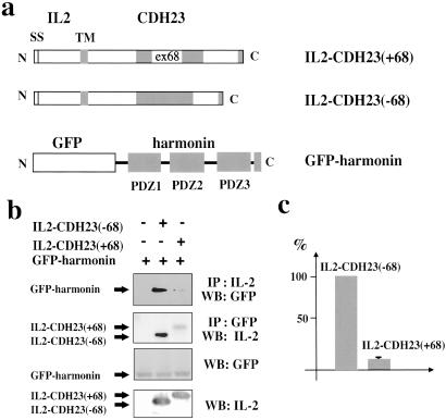
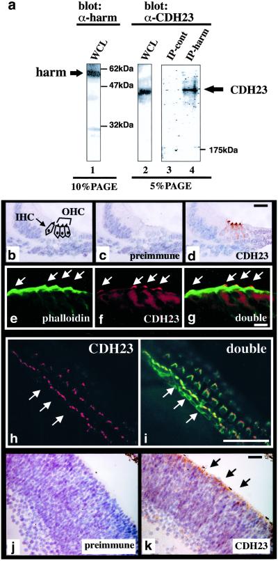
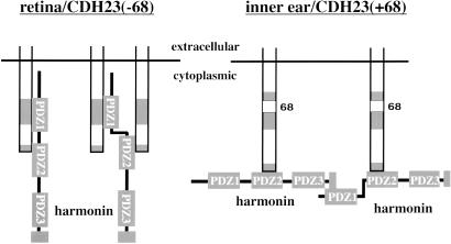
Usher syndrome type 1 (USH1) patients suffer from sensorineuronal deafness, vestibular dysfunction, and visual impairment. Several genetic loci have been linked to USH1, and four of the relevant genes have been identified. They encode the unconventional myosin VIIa, the PDZ-domain protein harmonin, and the putative adhesion receptors cadherin 23 (CDH23) and protocadherin 15 (PCDH15). We show here that CDH23 and harmonin form a protein complex. Two PDZ domains in harmonin interact with two complementary binding surfaces in the CDH23 cytoplasmic domain. One of the binding surfaces is disrupted by sequences encoded by an alternatively spliced CDH23 exon that is expressed in the ear, but not the retina. In the ear, CDH23 and harmonin are expressed in the stereocilia of hair cells, and in the retina within the photoreceptor cell layer. Because CDH23-deficient mice have splayed stereocilia, our data suggest that CDH23 and harmonin are part of a transmembrane complex that connects stereocilia into a bundle. Defects in the formation of this complex are predicted to disrupt stereocilia bundles and cause deafness in USH1 patients.


Figures

Fig 1.
Fig 1.
Hair cell diagram, protein–protein interaction domains in CDH23 and harmonin, and alternative splicing of CDH23. (a) A diagram of a hair cell (HC) is shown. The stereocilia are anchored at the cuticular plate (CP) to the hair cell body. They are connected to each other through extracellular linkages such as the tip link (TL), top connectors (TC), side links (SL), and ankle links (AL). (b) The cytoplasmic domain of CDH23 contains two putative PBIs. The C terminus of CDH23 fits the consensus sequence for class-I PBIs (30). An internal domain in the CDH23(−68) cytoplasmic domain shows homology to a PBI in Ril (35). In the CDH23(+68) isoform, 35 aa that are encoded by the alternatively spliced exon 68 are inserted into the Ril homology region. Gray boxes outline amino acids that are identical/conserved between CDH23 and Ril. Identical amino acids are indicated by an asterisk (*). (c) RT-PCR was conducted with RNA from adult mice with two sets of CDH23-specific primers (see b). The identity of the fragments was confirmed by DNA sequencing. The CDH23 isoform containing exon 68 was expressed in the inner ear but not in the brain or retina. MW, molecular weight marker. (d) Harmonin contains three PDZ domains (19, 20) and a class-I PBI. PDZ1 and PDZ2 have similarity to PDZ domains in PSD-95 (43, 44). PDZ3 shows similarity to a PDZ domain in p55 (38).
Fig 2.
Fig 2.
In yeast, harmonin interacts with harmonin and with CDH23. (a and b) Diagram of the harmonin and CDH23 fragments used in the two-hybrid assays. (c) Representative example of a yeast growth assay to measure harmonin–harmonin interactions. (Left) Growth in nonselective medium (control); (Right) growth in selective medium lacking histidine (selection). (d) Summary of the results establishing harmonin–harmonin interactions. (e) Representative example of a yeast growth assay to determine CDH23–harmonin interactions. (f) Summary of the results establishing CDH23–harmonin interaction. +, growth; −, no growth under selective pressure; n.d., not determined.
Fig 3.
Fig 3.
Exon 68-encoded amino acids affect CDH23–harmonin interactions in GST pull-down experiments. (a) Harmonin and its PDZ domains were expressed by in vitro transcription/translation in the presence of [35S]Met, resolved by SDS/PAGE, and visualized by autoradiography. (b) Purified GST-CDH23 fusion proteins were resolved on SDS-polyacrylamide gels and visualized by Coomassie staining. (c) Representative GST pull-down experiment. The input amount of GST-fusion proteins is shown (Lower). (d) The amount of harmonin that formed a complex with GST-CDH23 isoforms was quantified by scintillation counting and normalized against the input amount of GST-CDH23. The amount of harmonin interacting with GST-CDH23(−68) was set at 100%. Three experiments were performed, and the mean ± SD was determined. (e and f) Individual PDZ domains were used in GST pull-down experiments. The input proteins are shown in a. (e) PDZ2 was used for the GST pull-down experiments. (f) PDZ1 and PDZ2 were independently generated by in vitro translation and mixed.
Fig 4.
Fig 4.
Coimmunoprecipitation experiments from transfected cells. (a) Diagram of the fusion proteins (SS, signal sequence; TM, transmembrane domain). (b) Extracts from cells transfected to express the constructs shown in a were used for immunoprecipitation (IP) experiments with antibodies specific for IL-2 or GFP. Proteins were separated by SDS/PAGE and visualized by Western blotting (WB) with GFP or IL-2 antibodies. As a control, proteins were separated by SDS/PAGE without immunoprecipitation and visualized by Western blotting. (c) The amount of harmonin that coprecipitated with IL-2 antibodies was densitometrically quantified. Three independent experiments were performed, and harmonin levels recovered with CDH23(−68) were set at 100%. The mean ± SD is indicated.
Fig 5.
Fig 5.
CDH23–harmonin complex formation in vivo, and CDH23 expression in hair cells and the retina. (a) Whole-cell extracts (WCL) from P6 mouse brains were resolved on SDS-polyacrylamide gels directly (lanes 1 and 2), after immunoprecipitation with harmonin-specific antiserum (lane 4), or after a mock immunoprecipitation without antiserum (lane 3). Proteins were detected by Western blot with harmonin (lane 1) or CDH23-specific antiserum (lanes 2–4). (b–i) Analysis of CDH23 expression in the apical part of the cochlea at P0. (b) The section shows the position of the inner and outer hair cells (IHC, OHC). (c) No signal was detected with preimmune serum. (d) CDH23 is expressed in hair cell stereocilia. (eg) Immunofluorescence analysis: (e) F-actin, labeled with FITC-phalloidin, is present in the cuticular plate and stereocilia (arrows); (f) CDH23 is expressed in stereocilia only; (g) double exposure. (h) Immunofluorescence analysis of a P6 whole mount cochlea stained with CDH23-specific antiserum, followed by rhodamine-conjugated secondary antibody. CDH23 is expressed in stereocilia of inner and outer hair cells. The arrows point to the one row of inner hair cells. (i) Whole mounts were labeled with FITC-phalloidin to reveal F-actin. The double exposure shows localization of CDH23 and F-actin. (j and k) Sections of P0 retinas were stained with preimmune serum or with CDH23-specific serum. CDH23 expression is strongest in the photoreceptor layer (arrows). [Bars = 15 μm (bd), 5 μm (eg), 30 μm (h and i), and 20 μm (j and k).]
Fig 6.
Fig 6.
Model for CDH23–harmonin complex formation and its regulation by alternative splicing. On the basis of our data, we propose the following model. PDZ1 and PDZ2 of harmonin interact with the internal and C-terminal PBI of CDH23(−68), respectively, which could lead to binding of harmonin to one CDH23 molecule, or it could cross-link two receptors (Left). Insertion of the sequence encoded by exon 68 perturbs interactions between PDZ1 of harmonin and the internal PBI of CDH23(+68). This perturbation poises the complex for alternative interactions (Right). Notably, the PDZ1 domain of harmonin may be available to interact with other proteins when harmonin is bound to CDH23(+68), but not to CDH23(−68). Binding partners for PDZ1 include harmonin, leading to the formation of harmonin–harmonin oligomers. Because CDH23(+68) is expressed in the ear but not the retina, splicing may control the architecture and composition of CDH23–harmonin complexes in a tissue-specific manner.

Similar articles

Cited by

References

    1. Müller U. & Evans, A. L. (2001) Trends Cell Biol. 11, 334-342. - PubMed
    1. Steel K. P. & Kros, C. J. (2001) Nat. Genet. 27, 143-149. - PubMed
    1. Hone S. W. & Smith, R. J. (2001) Semin. Neonatol. 6, 531-541. - PubMed
    1. Richardson G. P., Bartolami, S. & Russell, I. J. (1990) J. Cell Biol. 110, 1055-1066. - PMC - PubMed
    1. Goodyear R. & Richardson, G. (1999) J. Neurosci. 19, 3761-3772. - PMC - PubMed

Publication types

LinkOut - more resources