

MRI of mouse models for gliomas shows similarities to humans and can be used to identify mice for preclinical trials
- PMID: 12407441
- PMCID: PMC1503661
- DOI: 10.1038/sj.neo.7900269
MRI of mouse models for gliomas shows similarities to humans and can be used to identify mice for preclinical trials
Abstract
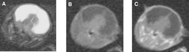
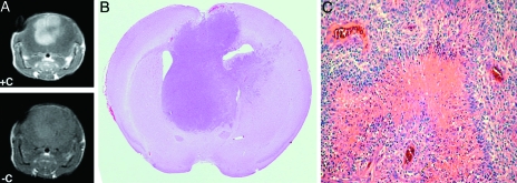
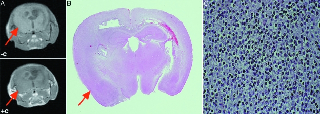
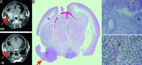
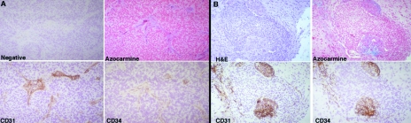
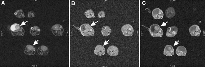
Magnetic resonance imaging (MRI) has been utilized for screening and detecting brain tumors in mice based upon their imaging characteristics appearance and their pattern of enhancement. Imaging of these tumors reveals many similarities to those observed in humans with identical pathology. Specifically, high-grade murine gliomas have histologic characteristics of glioblastoma multiforme (GBM) with contrast enhancement after intravenous administration of gadolinium diethylenetriamine pentaacetic acid (Gd-DTPA), implying disruption of the blood-brain barrier in these tumors. In contrast, low-grade murine oligodendrogliomas do not reveal contrast enhancement, similar to human tumors. MRI can be used to identify mice with brain neoplasms as inclusion criteria in preclinical trials.
Figures
Similar articles
-
Serial nonenhancing magnetic resonance imaging scans of high grade glioblastoma multiforme.J Natl Med Assoc. 1993 Feb;85(2):122-8. J Natl Med Assoc. 1993. PMID: 8382751 Free PMC article.
-
Combined activation of Ras and Akt in neural progenitors induces glioblastoma formation in mice.Nat Genet. 2000 May;25(1):55-7. doi: 10.1038/75596. Nat Genet. 2000. PMID: 10802656
-
Early versus late GD-DTPA MRI enhancement in experimental glioblastomas.J Magn Reson Imaging. 2011 Mar;33(3):550-6. doi: 10.1002/jmri.22472. J Magn Reson Imaging. 2011. PMID: 21563238
-
Radiological features of infantile glioblastoma and desmoplastic infantile tumors: British Columbia's Children's Hospital experience.J Neurosurg Pediatr. 2015 Aug;16(2):119-25. doi: 10.3171/2014.10.PEDS13634. Epub 2015 May 8. J Neurosurg Pediatr. 2015. PMID: 25955808 Review.
-
Basic principles of mathematical growth modeling applied to high-grade gliomas: A brief clinical review for clinicians.Neurol India. 2018 Nov-Dec;66(6):1575-1583. doi: 10.4103/0028-3886.246238. Neurol India. 2018. PMID: 30504543 Review.
Cited by
-
Modeling Adult Gliomas Using RCAS/t-va Technology.Transl Oncol. 2009 May;2(2):89-95. doi: 10.1593/tlo.09100. Transl Oncol. 2009. PMID: 19412424 Free PMC article.
-
Imaging in neurooncology.NeuroRx. 2005 Apr;2(2):333-47. doi: 10.1602/neurorx.2.2.333. NeuroRx. 2005. PMID: 15897954 Free PMC article. Review.
-
Multiple-mouse MRI with multiple arrays of receive coils.Magn Reson Med. 2010 Mar;63(3):803-10. doi: 10.1002/mrm.22236. Magn Reson Med. 2010. PMID: 20146352 Free PMC article.
-
Molecular imaging of a fluorescent antibody against epidermal growth factor receptor detects high-grade glioma.Sci Rep. 2021 Mar 11;11(1):5710. doi: 10.1038/s41598-021-84831-4. Sci Rep. 2021. PMID: 33707521 Free PMC article.
-
Integrin αvβ3-targeted IRDye 800CW near-infrared imaging of glioblastoma.Clin Cancer Res. 2012 Oct 15;18(20):5731-40. doi: 10.1158/1078-0432.CCR-12-0374. Epub 2012 Aug 22. Clin Cancer Res. 2012. PMID: 22914772 Free PMC article.
References
-
- Provenzale JM, Wang GR, Brenner T, Petrella JR, Sorenson AG. Comparison of permeability in high-grade and low-grade brain tumors using dynamic susceptibility contrast MR imaging. Am J Roentgenol. 2002;178:711–716. - PubMed
-
- Chenevert TL, McKeever PE, Ross BD. Monitoring early response of experimental brain tumors to therapy using diffusion magnetic resonance imaging, (1997) Clin Cancer Res. 1997;3:1457–1466. - PubMed
Publication types
MeSH terms
Substances
Grants and funding
LinkOut - more resources
Full Text Sources
Medical
Molecular Biology Databases