Comparative Study
. 2002 Nov-Dec;4(6):493-500.
doi: 10.1038/sj.neo.7900262.

Cooperation of hTERT, SV40 T antigen and oncogenic Ras in tumorigenesis: a cell transplantation model using bovine adrenocortical cells

Affiliations
Comparative Study

Cooperation of hTERT, SV40 T antigen and oncogenic Ras in tumorigenesis: a cell transplantation model using bovine adrenocortical cells

Michael Thomas et al. Neoplasia. 2002 Nov-Dec.

Abstract

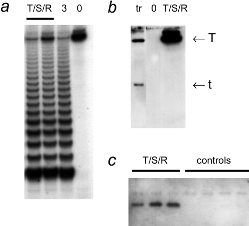
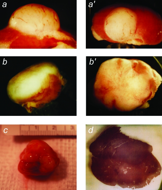
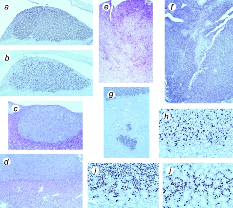
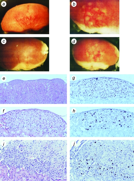
Expression of TERT, the reverse transcriptase component of telomerase, is necessary to convert normal human cells to cancer cells. Despite this, "telomerization" by hTERT does not appear to alter the normal properties of cells. In a cell transplantation model in which bovine adrenocortical cells form vascularized tissue structures beneath the kidney capsule in scid mice, telomerization does not perturb the functional tissue-forming capacity of the cells. This cell transplantation model was used to study the cooperation of hTERT with SV40 T antigen (SV40 TAg) and oncogenic Ras in tumorigenesis. Only cells expressing all three genes were tumorigenic; this required large T, but not small t, antigen. These cells produced a continuously expanding tissue mass; they were invasive with respect to adjacent organs and eventually destroyed the kidney. Cells expressing only hTERT or only Ras produced minimally altered tissues. In contrast, SV40 TAg alone produced noninvasive nodules beneath the kidney capsule that had high proliferation rates balanced by high rates of apoptosis. The use of cell transplantation techniques in a cell type that is able to form tissue structures with or without full neoplastic conversion allows the phenotypes produced by individual cooperating oncogenes to be observed.


Figures

Figure 1
Figure 1
Biochemical characterization of bovine adrenocortical cells transduced with hTERT, SV40 TAg, and Ras retroviruses. (a) Telomerase activity. TRAP assays were performed on extracts of hTERT→SV40 TAg→Ras cells (T/S/R; 0.25 and 0.5 µg of protein) and on an extract of 3T3 cells (“3”; 0.5 µg, positive control). “0” indicates the reaction performed with buffer only. (b) SV40 TAg. Western blot probed with an antibody that recognizes both large T and small t antigens. Extracts (100 µg of protein each) from hTERT→SV40 TAg→Ras cells and from clone 47 (Ref. [5]; cells transfected with hTERT and SV40 plasmids; labeled “tr”). (c) Ras. Extracts of separately grown cultures of hTERT→SV40 TAg→Ras cells and from separate batches of primary nongenetically modified cells (controls) were used for immunoprecipitation with an anti-Ras antibody, followed by Western blotting using an antibody against RasV12, as described in Materials and Methods section.
Figure 2
Figure 2
Macroscopic appearance of tumors produced from transplanted hTERT→SV40 TAg→Ras cells. After growth in culture, cells were transplanted under the kidney capsule of scid mice. Animals were sacrificed at intervals of 30 to 75 days following cell transplantation. The tumor masses found to have resulted from growth of the transplanted cells were photographed, together with the host animal kidney. (a,a′) Example of kidney and tissue mass removed from an animal at 30 days (magnification, x12, x8). (b,b′) Tissue mass removed at 45 days (magnification, x4, x6). (c) Tumor and kidney removed at 60 days (magnification, x2). (d) Gross appearance of a part of a similar tumor and kidney removed at 60 days and cut transversely (the tumor mass is the darker tissue above the kidney) (magnification, x8).
Figure 3
Figure 3
Histological appearance of tumors formed from hTERT→SV40 TAg→Ras cells, showing proliferation and invasive behavior. (a) Tumor mass above mouse kidney, removed at 30 days, stained with an antibody against SV40 TAg. (b) Serial section from same specimen stained for Ki-67 proliferation-specific antigen. (c) Hematoxylin and eosin-stained section of tumor mass in relation to kidney, removed at 30 days. (d) Hematoxylin and eosin-stained section of tumor mass above kidney, removed at 60 days. (e) Hematoxylin and eosin-stained section of late tumor, removed at 75 days, showing advanced invasion and destruction of mouse kidney. (f) Similar stage of tumor stained for SV40 TAg. (g) Part of kidney at 60 days, stained for SV40 TAg, showing detached mass of tumor cells within kidney. (h) Similar specimen showing border of kidney and tumor, stained for Ki-67 antigen (note that mouse Ki-67 is not recognized by this antibody). (i,j) Similar specimen, stained for SV40 TAg, showing invasion of tumor cells into kidney; note cells surrounding kidney tubules in (j). Magnification (a–d), x100; (e–g), x50; (h–j), x400.
Figure 4
Figure 4
Tissues formed from SV40 TAg cells and hTERT→SV40 TAg cells. Bovine adrenocortical cells were transduced with SV40 TAg retrovirus (a–h) or were successively infected with hTERT and SV40 TAg retroviruses (i,j). (a–d) Gross appearance of SV40 TAg tissues formed on surface of mouse kidney; four examples, removed from host animals at 45 to 60 days (magnification, x10, x20, x10, x10). (e) Hematoxylin and eosin-stained section of kidney showing transplant tissue beneath capsule as a series of small nodules (magnification, x40). (f) Enlarged view of one nodule (x200). (g) Section stained for SV40 TAg (x200). (h) Section stained for Ki-67 (x200). (i) Hematoxylin and eosin-stained section of hTERT→SV40 TAg tissue (x200). (j) hTERT→SV40 TAg tissue stained for Ki-67 (x200).
Figure 5
Figure 5
Cell death in tissue formed from SV40 TAg cells. Tissue was formed from bovine adrenocortical cells transduced with SV40 TAg retrovirus, or hTERT→SV40 TAg→Ras. Sections were used for ISOL detection of apoptotic cells. (a) Tissue formed from SV40 TAg cells. (b) Tissue formed from hTERT→SV40 TAg→Ras cells. Stained cells are marked with arrowheads. (b) shows the only two such cells found in an entire section of hTERT→SV40 TAg→Ras tumor. Magnification, x500.
Figure 6
Figure 6
Tissues formed from Ras and hTERT cells. Bovine adrenocortical cells were infected with retroviruses encoding either RasG12V (a–c) or hTERT (d,e). (a,b,d) Hematoxylin and eosin-stained sections; magnification, x40, x200, x150. (c,e) Proliferation visualized by staining with antibody against Ki-67 antigen; magnification, x200, x150.
Figure 7
Figure 7
Expression of Ras in tissues formed from bovine adrenocortical cells infected with RasG12V retrovirus; tissues stained with anti-Ras antibody. (a,b) Two examples of tissues formed from cells infected with RasG12V retrovirus only. (c) Tissue formed from hTERT→SV40 TAg→Ras cells. (d) Tissue formed from control bovine adrenocortical cells, not genetically modified; magnification, x200.

Similar articles

Cited by

References

    1. Greider CW. Telomeres and senescence: the history, the experiment, the future. Curr Biol. 1998;8:R178–R181. - PubMed
    1. Bodnar AG, Ouellette M, Frolkis M, Holt SE, Chiu CP, Morin GB, Harley CB, Shay JW, Lichsteiner S, Wright WE. Extension of life-span by introduction of telomerase into normal human cells. Science. 1998;279:349–352. - PubMed
    1. Wright WE, Shay JW. Cellular senescence as a tumor-protection mechanism: the essential role of counting. Curr Opin Genet Dev. 2001;11:98–103. - PubMed
    1. Lanza RP, Cibelli JB, Blackwell C, Cristofalo VJ, Francis MK, Baerlocher GM, Mak J, Schertzer M, Chavez EA, Sawyer N, Lansdorp PM, West MD. Extension of cell life-span and telomere length in animals cloned from senescent somatic cells. Science. 2000;288:665–669. - PubMed
    1. Thomas M, Yang L, Hornsby PJ. Formation of normal functional tissue from transplanted adrenocortical cells expressing telomerase reverse transcriptase. Nat Biotechnol. 2000;18:39–42. - PubMed

Publication types

MeSH terms