

Real-time PCR method for detection of Encephalitozoon intestinalis from stool specimens
- PMID: 12409353
- PMCID: PMC139654
- DOI: 10.1128/JCM.40.11.3922-3928.2002
Real-time PCR method for detection of Encephalitozoon intestinalis from stool specimens
Abstract
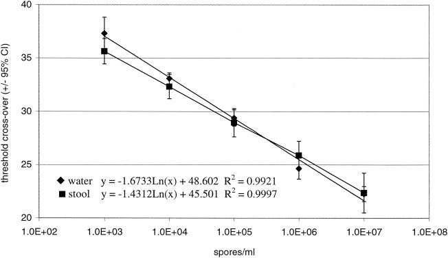
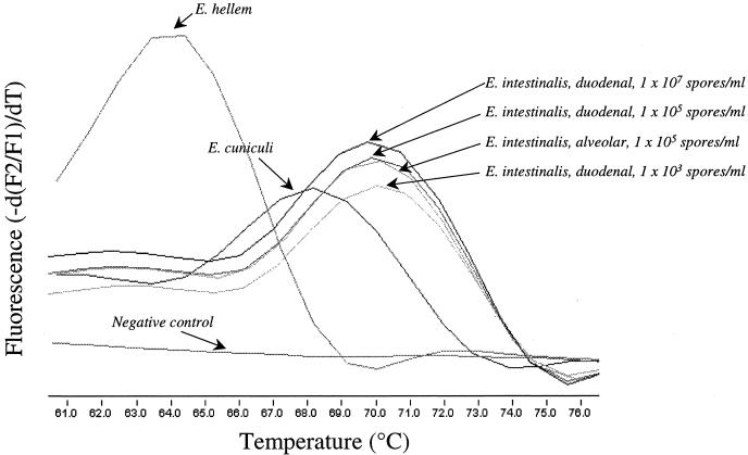
The prevalence of microsporidiosis is likely underestimated due to the labor-intensive, insensitive, and nonspecific clinical laboratory methods used for the diagnosis of this disease. A real-time PCR assay was designed to assess DNA extraction methods and to detect three Encephalitozoon species in feces. Modifications of the MagNA Pure LC DNA isolation kit protocol (Roche Applied Sciences, Indianapolis, Ind.) were compared by using the automated MagNA Pure LC instrument (Roche) and fecal specimens spiked with Encephalitozoon intestinalis spores. Extracted DNA was amplified by the LightCycler (Roche) PCR assay. Assay sensitivity, reproducibility, and efficiency were assessed by comparing threshold crossover values achieved with different extraction and storage conditions (fresh, refrigerated, frozen, and preserved specimens). Optimal extraction conditions were achieved by using a commercial buffer, tissue lysis buffer (Roche), as the specimen diluent. LightCycler PCR results were compared to those obtained from routine stool microscopy with trichrome blue stain. The lower limit of detection for the LightCycler PCR assay varied by storage conditions from 10(2) to 10(4) spores/ml of feces, a value which represented a significant improvement over that achieved by staining (> or =1.0 x 10(6) spores/ml). Melting temperature analysis of the amplicons allowed for the differentiation of three Encephalitozoon species (E. intestinalis, E. cuniculi, and E. hellem). The assay is readily adaptable to the clinical laboratory and represents the first real-time PCR assay designed to detect Encephalitozoon species.
Figures
References
-
- Akane, A., K. Matsubara, H. Nakamura, S. Takahashi, and K. Kimura. 1994. Identification of the heme compound copurified with deoxyribonucleic acid (DNA) from bloodstains, a major inhibitor of polymerase chain reaction (PCR) amplification. J. Forensic Sci. 39:362-372. - PubMed
-
- Ali, M. S., L. A. Mahmoud, B. E. Abaza, and M. A. Ramadan. 2000. Intestinal spore-forming protozoa among patients suffering from chronic renal failure. J. Egypt. Soc. Parasitol. 30:93-100. - PubMed
Publication types
MeSH terms
Substances
LinkOut - more resources
Full Text Sources
Other Literature Sources
Medical
