. 2002 Nov;40(11):4105-13.
doi: 10.1128/JCM.40.11.4105-4113.2002.

Human cytomegalovirus DNA in plasma and serum specimens of renal transplant recipients is highly fragmented

Affiliations

Human cytomegalovirus DNA in plasma and serum specimens of renal transplant recipients is highly fragmented

René Boom et al. J Clin Microbiol. 2002 Nov.

Abstract

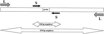
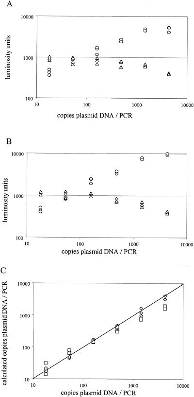
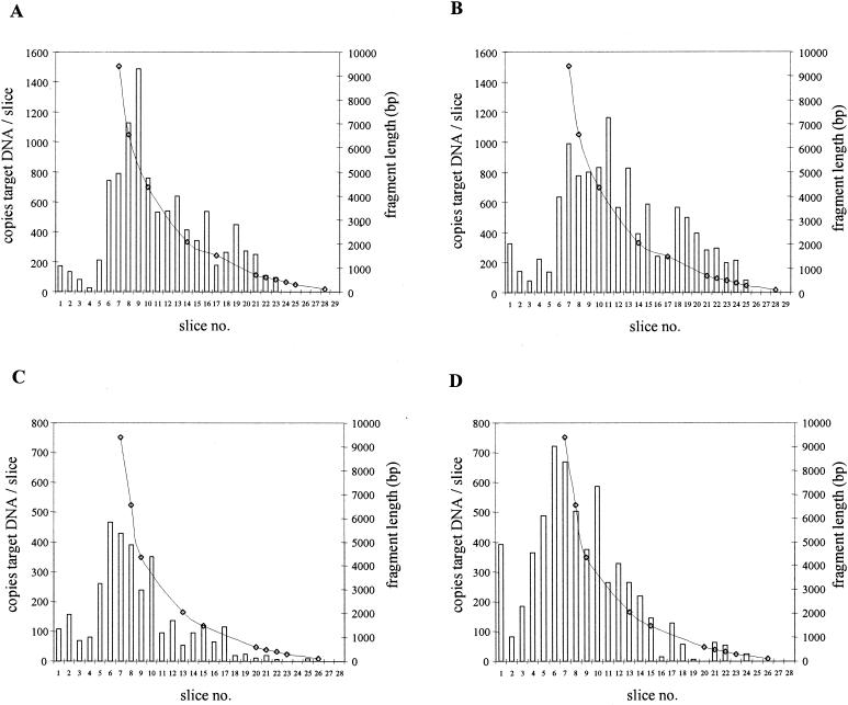
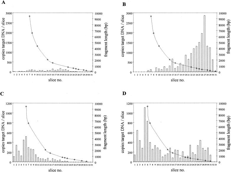
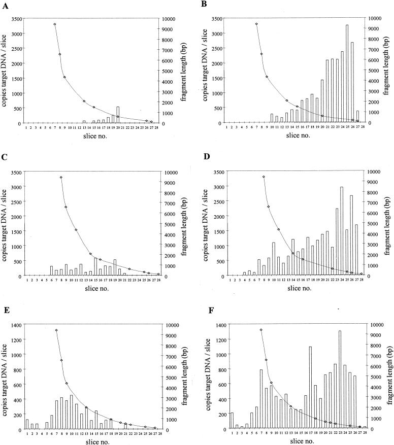
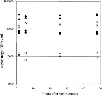
Quantitation of cytomegalovirus (CMV) DNA in plasma and serum by PCR is increasingly used to identify patients at risk for developing CMV disease and to monitor the efficacy of antiviral therapy. Although CMV DNA levels are generally interpreted as viral loads, the exact nature of the viral DNA in these specimens is unknown. We studied the state of CMV DNA in plasma and serum specimens obtained from three renal transplant recipients at peak viral DNA levels during primary CMV infection. For this purpose, DNA isolated from these specimens was fractionated by size, and CMV DNA levels in the resulting DNA fractions were measured by quantitative PCR targeted at large (578-bp) and small (134-bp) amplicons. These experiments showed that the molecular sizes of DNA fragments from which CMV DNA is amplified were small (<2,000 bp), indicating that CMV DNA in plasma and serum is highly fragmented. Furthermore, CMV DNA levels were consistently higher when targeted at the smaller amplicon, providing additional evidence for the fragmentation of viral DNA. In conclusion, the first results with three patients have shown that CMV DNA in plasma and serum is highly fragmented and does not necessarily reflect the amount of infectious virus. These observations have potential consequences for understanding CMV pathogenesis and interpreting CMV DNA levels in individual patient management.


Figures

FIG. 1.
FIG. 1.
Schematic representation (not to scale) of primer pair L and primer pair S, used for amplification of 578- and 138-bp amplicons, respectively. The 25-bp region in which the CMV-specific probe and the IC-specific probe are located is indicated. Except for the probe region, CMV and IC sequences are similar. For details, see Materials and Methods.
FIG. 2.
FIG. 2.
Validation of QPCR for 138- and 578-bp amplicons. (A and B) A serial threefold dilution of plasmid pES was subjected in triplicate to QPCR with the S (A) and L (B) primer pairs. Raw data (in luminosity units) obtained after hybridization with the IC-specific probe (triangles) and the CMV-specific probe (circles) are given. (C) From the raw data, the number of copies target DNA was calculated from the ratio of CMV-specific and IC-specific signals. Diamonds, 578-bp amplicon; squares, 138-bp amplicon.
FIG. 3.
FIG. 3.
Size distributions of PCR targets for CMV DNA isolated from purified CMV virions (A and B) and from Rat-9G cells (C and D) for the 578-bp (A and C) and 138-bp (B and D) amplicons. Slice 1 represents the slot to which DNA was applied in the agarose gel. Bars indicate the number of copies of CMV DNA molecules per agarose slice. Lines represent the lengths of the DNA fragments from which CMV DNA was amplified.
FIG. 4.
FIG. 4.
Size distributions of CMV DNA targets in plasma (A and B) and in whole blood (C and D) from a renal transplant recipient (recipient A) for the 578-bp (A and C) and 138-bp (B and D) amplicons. The amounts of DNA applied to the gel corresponded to 12.5 μl of whole blood and 50 μl of plasma. Slice 1 represents the slot in the agarose gel. Bars indicate the number of copies of CMV DNA molecules per agarose slice. Lines represent the lengths of the DNA fragments from which CMV DNA was amplified.
FIG. 5.
FIG. 5.
Size distributions of CMV DNA targets in serum (A and B), EDTA-plasma (C and D), and whole blood (E and F) from a renal transplant recipient (recipient B) for the 578-bp (A, C, and E) and 138-bp (B, D, and F) amplicons. The amounts of DNA applied to the gel corresponded to 12.5 μl of whole blood and 50 μl of plasma or serum. Slice 1 represents the slot in the agarose gel. Bars indicate the number of copies of CMV DNA molecules per agarose slice. Lines represent the lengths of the DNA fragments from which CMV DNA was amplified.
FIG. 6.
FIG. 6.
Stability of CMV DNA targets in whole blood and plasma prepared at different time points after venipuncture of recipient C. Results are from duplicate extractions. Triangles, whole blood; circles, EDTA-plasma. Open symbols, loads obtained for the 578-bp amplicon; closed symbols, loads obtained for the 138-bp amplicon.

References

    1. Aaij, C., and P. Borst. 1972. The gel electrophoresis of DNA. Biochim. Biophys. Acta 269:192-200. - PubMed
    1. Akrigg, A., G. W.G. Wilkinson, and J. D. Oram. 1985. The structure of the major immediate early gene of human cytomegalovirus strain AD169. Virus Res. 2:107-121. - PubMed
    1. Boeckh, M., and G. Boivin. 1998. Quantitation of cytomegalovirus: methodologic aspects and clinical applications. Clin. Microbiol. Rev. 11:533-554. - PMC - PubMed
    1. Boivin, G., J. Handfield, E. Toma, G. Murray, R. Lalonde, and G. Bergeron. 1998. Comparative evaluation of the cytomegalovirus DNA load in polymorphonuclear leukocytes and plasma of human immunodeficiency virus-infected subjects. J. Infect. Dis. 177:355-360. - PubMed
    1. Boivin, G., J. Handfield, E. Toma, R. Lalonde, and M. G. Bergeron. 1999. Expression of the late cytomegalovirus (CMV) pp150 transcript in leukocytes of AIDS patients is associated with a high viral load in leukocyte and presence of CMV DNA in plasma. J. Infect. Dis. 179:1101-1107. - PubMed

MeSH terms