

Development of a peptide-mediated capture PCR for detection of Mycobacterium avium subsp. paratuberculosis in milk
- PMID: 12409405
- PMCID: PMC139712
- DOI: 10.1128/JCM.40.11.4244-4250.2002
Development of a peptide-mediated capture PCR for detection of Mycobacterium avium subsp. paratuberculosis in milk
Abstract
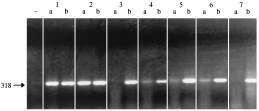
Based on phage display technology, a peptide-mediated magnetic separation technique was developed to facilitate selective isolation of Mycobacterium avium subsp. paratuberculosis (M. paratuberculosis) from bulk milk of naturally infected dairy herds. Nine recombinant bacteriophages binding to M. paratuberculosis were isolated from a commercial phage-peptide library encoding random 12-mer peptides. Nucleotide sequencing revealed the deduced sequence of the binding peptides. One peptide with the sequence NYVIHDVPRHPA, designated aMP3, was chemically synthesized with an amino-terminal biotin residue attached via an amino-hexacarbonic acid spacer molecule. Paramagnetic beads coated with the phage or with peptide aMP3 enabled the capture of M. paratuberculosis from milk. Combining this peptide-mediated magnetic separation with an ISMav2-based PCR allowed the detection of M. paratuberculosis in artificially spiked milk down to a concentration of 10(1) ml(-1). Experiments using milk from naturally infected cows and bulk milk samples from infected herds demonstrated that the peptide-mediated capture PCR is sufficiently sensitive to detect single strong shedders in pooled milk samples. The method, for the first time, applies phage display technology to microbial diagnostics and has potential value as a completely standardizable tool for the routine M. paratuberculosis screening of bulk milk samples at acceptable costs.
Figures
Similar articles
-
Maximizing capture efficiency and specificity of magnetic separation for Mycobacterium avium subsp. paratuberculosis cells.Appl Environ Microbiol. 2010 Nov;76(22):7550-8. doi: 10.1128/AEM.01432-10. Epub 2010 Sep 17. Appl Environ Microbiol. 2010. PMID: 20851966 Free PMC article.
-
Peptide aMptD-mediated capture PCR for detection of Mycobacterium avium subsp. paratuberculosis in bulk milk samples.Appl Environ Microbiol. 2006 Aug;72(8):5150-8. doi: 10.1128/AEM.00590-06. Appl Environ Microbiol. 2006. PMID: 16885259 Free PMC article.
-
Automated high-throughput immunomagnetic separation-PCR for detection of Mycobacterium avium subsp. paratuberculosis in bovine milk.Int J Food Microbiol. 2006 Aug 1;110(3):201-8. doi: 10.1016/j.ijfoodmicro.2006.01.042. Epub 2006 Jul 11. Int J Food Microbiol. 2006. PMID: 16814891
-
Mycobacterium paratuberculosis detection in cow's milk in Argentina by immunomagnetic separation-PCR.Braz J Microbiol. 2016 Apr-Jun;47(2):506-12. doi: 10.1016/j.bjm.2016.01.013. Epub 2016 Mar 2. Braz J Microbiol. 2016. PMID: 26991290 Free PMC article.
-
Magnetic Separation Methods for the Detection of Mycobacterium avium subsp. paratuberculosis in Various Types of Matrices: A Review.Biomed Res Int. 2017;2017:5869854. doi: 10.1155/2017/5869854. Epub 2017 May 31. Biomed Res Int. 2017. PMID: 28642876 Free PMC article. Review.
Cited by
-
Maximizing capture efficiency and specificity of magnetic separation for Mycobacterium avium subsp. paratuberculosis cells.Appl Environ Microbiol. 2010 Nov;76(22):7550-8. doi: 10.1128/AEM.01432-10. Epub 2010 Sep 17. Appl Environ Microbiol. 2010. PMID: 20851966 Free PMC article.
-
Detection of Mycobacterium avium subsp. paratuberculosis by a sonicate immunoassay based on surface-enhanced Raman scattering.Clin Vaccine Immunol. 2008 Feb;15(2):227-34. doi: 10.1128/CVI.00334-07. Epub 2007 Dec 12. Clin Vaccine Immunol. 2008. PMID: 18077613 Free PMC article.
-
Production and evaluation of antibodies and phage display-derived peptide ligands for immunomagnetic separation of Mycobacterium bovis.J Clin Microbiol. 2012 May;50(5):1598-605. doi: 10.1128/JCM.05747-11. Epub 2012 Feb 8. J Clin Microbiol. 2012. PMID: 22322353 Free PMC article.
-
Characterization of genetic differences between Mycobacterium avium subsp. paratuberculosis type I and type II isolates.J Clin Microbiol. 2003 Nov;41(11):5215-23. doi: 10.1128/JCM.41.11.5215-5223.2003. J Clin Microbiol. 2003. PMID: 14605167 Free PMC article.
-
Phages harboring specific peptides that recognize the N protein of the porcine reproductive and respiratory syndrome virus distinguish the virus from other viruses.J Clin Microbiol. 2010 May;48(5):1875-81. doi: 10.1128/JCM.01707-09. Epub 2010 Mar 17. J Clin Microbiol. 2010. PMID: 20237096 Free PMC article.
References
-
- Adda, C. G., R. F. Anders, L. Tilley, and M. Foley. 2002. Random sequence libraries displayed on phage: identification of biologically important molecules. Comb. Chem. High Throughput Screen 5:1-14. - PubMed
-
- Bakker, D., P. T. Willemsen, and F. G. van Zijderveld. 2000. Paratuberculosis recognized as a problem at last: a review. Vet. Q. 22:200-204. - PubMed
-
- Beard, P. M., K. Stevenson, A. Pirie, K. Rudge, D. Buxton, S. M. Rhind, M. C. Sinclair, L. A. Wildblood, D. G. Jones, and J. M. Sharp. 2001. Experimental paratuberculosis in calves following inoculation with a rabbit isolate of Mycobacterium avium subsp. paratuberculosis. J. Clin. Microbiol. 39:3080-3084. - PMC - PubMed
-
- Bickley, J., J. K. Short, D. G. McDowell, and H. C. Parkes. 1996. Polymerase chain reaction (PCR) detection of Listeria monocytogenes in diluted milk and reversal of PCR inhibition caused by calcium ions. Lett. Appl. Microbiol. 22:153-158. - PubMed
Publication types
MeSH terms
Substances
LinkOut - more resources
Full Text Sources
Other Literature Sources
Molecular Biology Databases
Miscellaneous
