

Assessment of the nasopharyngeal bacterial flora of rhesus macaques: moraxella, Neisseria, haemophilus, and other genera
- PMID: 12409426
- PMCID: PMC139662
- DOI: 10.1128/JCM.40.11.4340-4342.2002
Assessment of the nasopharyngeal bacterial flora of rhesus macaques: moraxella, Neisseria, haemophilus, and other genera
Abstract
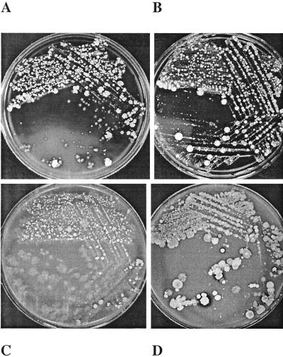
The nasopharyngeal bacterial flora of healthy rhesus macaques was surveyed for the presence of Neisseria and Haemophilus species, as well as Moraxella catarrhalis. M. catarrhalis was found both in healthy rhesus macaques and in possibly immunocompromised rhesus macaques. Several Haemophilus spp. that are part of the normal nasopharyngeal bacterial flora of humans were found in many animals; these Haemophilus species included H. parahaemolyticus, H. segnis, and H. parainfluenzae. While Haemophilus influenzae was not identified, it is possible that the identification of H. influenzae types may have been thwarted by the growth of less fastidious species. A number of animals harbored Neisseria spp. such as N. sicca. However, Neisseria meningitidis was not found. In summary, it appears as though the rhesus macaque may be used as a model for M. catarrhalis infections. Moreover, in view of the susceptibility of macaques to organisms of the Haemophilus and Neisseria genera, it is possible that these animals may also accurately model nontypeable H. influenzae and N. meningitidis infections.
Figures
References
-
- Hirsch, V. M., and J. D. Lifson. 2000. Simian immunodeficiency virus infection of monkeys as a model system for the study of AIDS pathogenesis, treatment, and prevention. Adv. Pharmacol. 49:437-477. - PubMed
Publication types
MeSH terms
Grants and funding
LinkOut - more resources
Full Text Sources
Other Literature Sources
Miscellaneous
