. 2002 Nov 1;30(21):4626-33.
doi: 10.1093/nar/gkf602.

Human mitochondrial DNA with large deletions repopulates organelles faster than full-length genomes under relaxed copy number control

Affiliations

Human mitochondrial DNA with large deletions repopulates organelles faster than full-length genomes under relaxed copy number control

Francisca Diaz et al. Nucleic Acids Res. .

Abstract

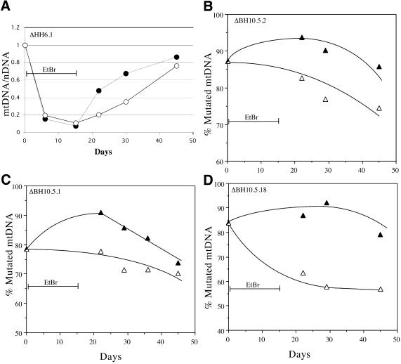
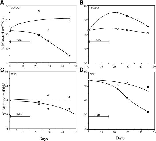
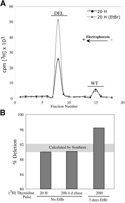
Partially-deleted mitochondrial DNA (DeltamtDNA) accumulates during aging of postmitotic tissues. This accumulation has been linked to decreased metabolic activity, increased reactive oxygen species formation and the aging process. Taking advantage of cell lines with heteroplasmic mtDNA mutations, we showed that, after severe mtDNA depletion, organelles are quickly and predominantly repopulated with DeltamtDNA, whereas repopulation with the wild-type counterpart is slower. This behavior was not observed for full-length genomes with pathogenic point mutations. The faster repopulation of smaller molecules was supported by metabolic labeling of mtDNA with [3H]thymidine during relaxed copy number control conditions. We also showed that hybrid cells containing two defective mtDNA haplotypes tend to retain the smaller one as they adjust their normal mtDNA copy number. Taken together, our results indicate that, under relaxed copy number control, DeltamtDNAs repopulate mitochondria more efficiently than full-length genomes.


Figures

Figure 1
Figure 1
Characterization of cell lines with mtDNA mutations used in this study. (A) Diagram illustrating the location of two point mutations, two large deletions and a 4 bp deletion in the apocytochrome b gene in the human mtDNA. (B) Southern blot showing the heteroplasmic mtDNA deletions in some of the cell lines used. Total DNA extracted from the different cell lines was digested with PvuII and analyzed by Southern hybridization to a probe corresponding to the ND1 gene. (C) PCR/RFLP analyses of the two pathogenic point mutations and PCR/LP of the apocytochrome b 4 bp deletion (A). The cell lines with tRNA mutations used in this study were heteroplasmic. Cell lines harboring homoplasmic levels of apocytochrome b 4 bp deletion, a 7.5 kb deletion and wild-type genomes were used in the hybrid experiments described below.
Figure 1
Figure 1
Characterization of cell lines with mtDNA mutations used in this study. (A) Diagram illustrating the location of two point mutations, two large deletions and a 4 bp deletion in the apocytochrome b gene in the human mtDNA. (B) Southern blot showing the heteroplasmic mtDNA deletions in some of the cell lines used. Total DNA extracted from the different cell lines was digested with PvuII and analyzed by Southern hybridization to a probe corresponding to the ND1 gene. (C) PCR/RFLP analyses of the two pathogenic point mutations and PCR/LP of the apocytochrome b 4 bp deletion (A). The cell lines with tRNA mutations used in this study were heteroplasmic. Cell lines harboring homoplasmic levels of apocytochrome b 4 bp deletion, a 7.5 kb deletion and wild-type genomes were used in the hybrid experiments described below.
Figure 2
Figure 2
ΔmtDNA heteroplasmy fluctuations after induced mtDNA depletion. (A) An osteosarcoma cell line containing high levels of a ΔmtDNA was treated with 50 ng/ml EtBr for 15 days and allowed to recover for 30 additional days. Cells were harvested at the indicated times and their DNA purified and analyzed for relative levels of mtDNA (mutated or wild-type, closed and open circles, respectively) / nDNA as described in Materials and Methods. (BD) Three cell lines harboring ΔmtDNA were subjected to a 15 day EtBr treatment followed by a 30 day recovery period. Parallel cultures growing in the same medium, but without EtBr were also analyzed to assess heteroplasmy fluctuations related to time in culture. Total DNA extracted at different times was analyzed by Southern blot and the percentage mutated mtDNA represented as filled triangles in the EtBr treated series and as open triangles in the control series. The mtDNA levels during EtBr treatment (days 7 and 15) were very low and did not allow a reliable estimation of heteroplasmy. There was a reduction in the percentage mutated mtDNA during normal growth conditions, but EtBr treatment was consistently associated with an increase in percentage mutated mtDNA immediately after the treatment.
Figure 3
Figure 3
Point mutant mtDNA heteroplasmy fluctuations after induced mtDNA depletion. Four cell lines harboring two different pathogenic mtDNA point mutations in tRNA genes were subjected to a 15 day EtBr treatment followed by a 30 day recovery period. Parallel cultures growing in the same medium, but without EtBr were performed to assess heteroplasmy fluctuations during time in culture. Total DNA was extracted at different times and analyzed by PCR/RFLP. The percentage mutated mtDNA is represented as filled circles in the EtBr treated series and as open circles in the control series (AD). With the exception of one cell line (B), the percentage of point mutated mtDNA either did not change significantly or decreased from pretreatment levels. The treatment with EtBr did not seem to alter this natural trend.
Figure 4
Figure 4
Metabolic labeling of mtDNA with [3H]thymidine. The cell line ΔDH2.1 was incubated with 20 µCi/ml of [3H]thymidine for the times indicated. In one group, cells were pretreated with EtBr for 3 days, allowed to recover without the drug for 24 h and labeled with [3H]thymidine for the periods indicated. mtDNA from labeled cells was purified from mitochondria isolated by N2 cavitation as described in Materials and Methods. mtDNA was digested with PvuII and separated by electrophoresis on a 0.8% agarose gel. The gel lane was sliced and the gel fractions analyzed in a scintillation counter (A). Percentage mutated mtDNA was calculated from the incorporated 3H after correction for the number of thymidines in each molecule. (B) Comparison of these values with the percentage mutation obtained from Southern blots (grey horizontal area).
Figure 5
Figure 5
Heteroplasmy levels of somatic hybrids generated without selection for OXPHOS function. Three different cell lines were fused to each other as depicted in (A). One contained wild-type mtDNA and a neomycin-resistance nuclear marker (N), a second was homoplasmic for a mtDNA 7.5 kb deletion containing a zeocin-resistance nuclear marker (Z) and a third was homoplasmic for a pathogenic 4 bp deletion in cytochrome b gene containing a puromycin-resistance nuclear marker (P). Fusion products were grown in high glucose media containing two of the respective selection drugs (i.e. G418, zeocin or puromycin) and supplemented with uridine. Surviving clones were isolated and their DNA analyzed by Southern blot as described in Materials and Methods. (B) Analysis of the cytochrome b 4 bp deletion in hybrids of PN cell lines. A 32P-labeled PCR amplicon, corresponding to the 5′ end of the apocytochrome b gene, was separated by electrophoresis on a 6% denaturing polyacrylamide gel and analyzed in a phosphoimager. (C) Phosphoimager signal from a Southern blot of DNA extracted from the hybrids between cells containing the 7.5 kb deletion and wt (NZ lines) or the 4 bp deletion in apocytochrome b (ZP lines).

References

    1. Shadel G.S. and Clayton,D.A. (1997) Mitochondrial DNA maintenance in vertebrates. Annu. Rev. Biochem., 66, 409–435. - PubMed
    1. Moraes C.T. (2001) What regulates mitochondrial DNA copy number in animal cells? Trends Genet., 17, 199–205. - PubMed
    1. Wallace D.C. (1997) Mitochondrial DNA in aging and disease. Sci. Am., 277, 40–47. - PubMed
    1. Raha S. and Robinson,B.H. (2000) Mitochondria, oxygen free radicals, disease and ageing. Trends Biochem. Sci., 25, 502–508. - PubMed
    1. Corral-Debrinski M., Horton,T., Lott,M.T., Shoffner,J.M., Beal,M.F. and Wallace,D.C. (1992) Mitochondrial DNA deletions in human brain: regional variability and increase with advanced age. Nature Genet., 2, 324–329. - PubMed

Publication types