. 2002 Nov 12;99(23):14716-21.
doi: 10.1073/pnas.232177599. Epub 2002 Oct 30.

Confirmation by FRET in individual living cells of the absence of significant amyloid beta -mediated caspase 8 activation

Affiliations

Confirmation by FRET in individual living cells of the absence of significant amyloid beta -mediated caspase 8 activation

Reiko Onuki et al. Proc Natl Acad Sci U S A. .

Abstract

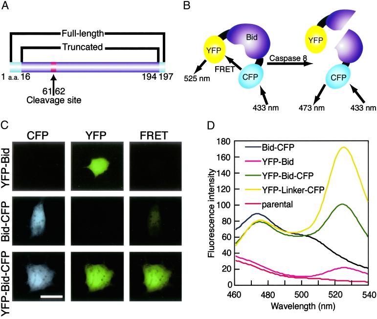
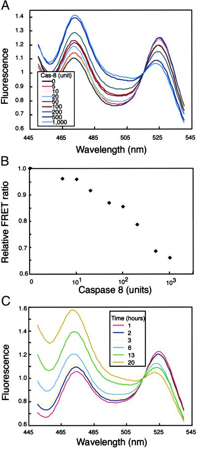
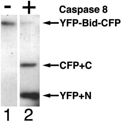
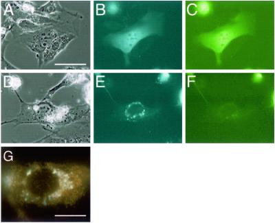
When cells are exposed to death-inducing molecules such as tumor necrosis factor-alpha or Fas, caspase 8 is activated and cleaves an apoptotic facilitator, Bid, that is a member of the Bcl-2 family. After additional modification, the C-terminal moiety of Bid is translocated to the mitochondria and induces the release of cytochrome c into the cytoplasm. In an attempt to directly observe the cleavage of Bid and the following events in living cells, we constructed a vector that encoded Bid fused with yellow fluorescent protein (YFP) and cyan fluorescent protein (CFP) (YFP-Bid-CFP). On expression of YFP-Bid-CFP in mammalian cells, we were able to observe the efficient transfer of energy from excited CFP to YFP within the YFP-Bid-CFP molecule and, importantly, the fusion protein YFP-Bid-CFP was fully functional in cells. When YFP-Bid-CFP was cleaved by caspase 8, on activation by anti-Fas Abs but not by Abeta or tunicamycin, no such transfer of energy was detected. To our knowledge, this is the first report of (i) visualization of the activation of Bid by proteolytic cleavage, with direct observation of the cleavage of YFP-Bid-CFP in the cytoplasm and subsequent translocation of the cleaved Bid to mitochondria and (ii) the absence of Abeta- or tunicamycin-mediated significant activation of caspase 8 in individual living cells.


Figures

Fig 1.
Fig 1.
(A) Schematic representation of the domain structure of CFP-Bid-YFP. YFP and CFP were connected to Bid (residues 16–194) by flexible spacers that consisted of three glycine residues each to generate CFP-Bid-YFP. The arrow between residues 61 and 62 indicates the site of cleavage by caspase 8. (B) In our FRET system, excitation of CFP at 433 nm should result in emission at 473 nm if no YFP is in close proximity. In the YFP-Bid-CFP, energy should be transferred from excited CFP to YFP, with resulting emission at 525 nm. When the fusion protein is cleaved by caspase 8, energy can no longer be transferred from excited CFP to YFP. (C) Fluorescence images of COS7 cells that expressed the indicated fusion proteins (y axis). Images were acquired by using FRET, CFP, and YFP filters (x axis). (Bar = 20 μm.) (D) Fluorescence spectra of extracts of cells that expressed various fusion proteins. The amount of total protein for each sample was adjusted to 1.4 mg/ml. The extracts of cells expressing YFP-Bid (pink line), Bid-CFP (black Line), YFP-Bid-CFP (green line), YFP-linker-CFP (yellow line), and parental cells (red line) were excited at 433 nm and emission spectra were recorded and normalized at 450 nm.
Fig 2.
Fig 2.
(A) Results of spectrofluorometric analysis of the cleavage of YFP-Bid-CFP in cell extracts by caspase 8. Extracts were incubated with increasing amounts of caspase 8 (Cas-8) for 22 h at 25°C, and then spectra were recorded after excitation at 433 nm. (B) The ratio of fluorescence from YFP (525 nm) to that from CFP (473 nm) was calculated for each concentration of caspase 8 in the reaction mixture. The relative FRET ratio was defined as the ratio of values of YFP fluorescence/CFP fluorescence measured before and after incubation with caspase 8. (C) Time-dependent cleavage of YFP-Bid-CFP, as demonstrated by spectrofluorometric analysis.
Fig 3.
Fig 3.
Western blotting analysis, showing the cleavage of YFP-Bid-CFP by caspase 8. Lane 1, control (no caspase 8) and lane 2, extract of COS 7 cells incubated with 1,000 units of caspase 8 for 22 h at 25°C. Extracts were separated by SDS/PAGE on a 10% polyacrylamide gel and subjected to Western blotting with an anti-GFP Ab. The upper arrow indicates YFP-Bid-CFP. The middle arrow indicates C-terminal Bid-CFP-fused protein, and the bottom arrow indicates YFP-N-terminal Bid-fused protein.
Fig 4.
Fig 4.
Subcellular localization of YFP-Bid-CFP. NIH 3T3 cells were transfected with pFRET-Bid and incubated for 52 h. Cells on DG were exposed to tumor necrosis factor-α plus cycloheximide for 3 h. Controls were not exposed to these agents (AC). After incubation, cells were observed by fluorescence microscopy. Phase-contrast images (A and D), images of CFP fluorescence (B and E), and images of YFP fluorescence (C and F). (Bar = 20 μm in AF.) The subcellular localization of truncated Bid (cyan fluorescence) and mitochondria (orange fluorescence) was assessed by fluorescence microscopy (G). (Bar = 10 μm.)
Fig 5.
Fig 5.
Fluorescence images of YFP-Bid-CFP and YFP-linker-CFP in cells treated with various inducers of apoptosis. (A) SK-N-SH cells were stably transfected with pFRET-Bid or pFRET-linker, and images were recorded under phase-contrast conditions (phase) and with FRET, CFP, and YFP filters. Linker refers to YFP-linker-CFP and Bid refers to YFP-Bid-CFP. YFP-linker-CFP and YFP-Bid-CFP diffused uniformly throughout the cells. (Bar = 20 μm.) (B) Cells were induced to undergo apoptosis by anti-Fas Ab (Fas), Tm, or Aβ. After treatment, images were recorded as indicated above. Apoptosis-induced cells were also observed under higher (B) and lower (C) magnification. (Bar = 10 μm.)

Similar articles

Cited by

References

    1. Li H. & Yuan, J. (1999) Curr. Opin. Cell Biol. 11, 261-266. - PubMed
    1. Korsmeyer S. J., Wei, M. C., Saito, M., Weiler, S., Oh, K. & Schlesinger, P. H. (2000) Cell Death Differ. 7, 1166-1173. - PubMed
    1. Wang K., Yin, X. M., Chao, D. T., Milliman, C. L. & Korsmeyer, S. J. (1996) Genes Dev. 10, 2859-2869. - PubMed
    1. Wei M. C., Lindsten, T., Mootha, V. K., Weiler, S., Gross, A., Ashiya, M., Thompson, C. B. & Korsmeyer, S. J. (2000) Genes Dev. 14, 2060-2071. - PMC - PubMed
    1. Gross A., Yin, X. M., Wang, K., Wei, M. C., Jockel, J., Milliman, C., Erdjument-Bromage, H., Tempst, P. & Korsmeyer, S. J. (1999) J. Biol. Chem. 274, 1156-1163. - PubMed

Publication types

MeSH terms