. 2002 Nov 1;21(21):5599-610.
doi: 10.1093/emboj/cdf566.

Structural basis for the oxidation of thiosulfate by a sulfur cycle enzyme

Affiliations

Structural basis for the oxidation of thiosulfate by a sulfur cycle enzyme

Vicki A Bamford et al. EMBO J. .

Abstract

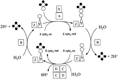
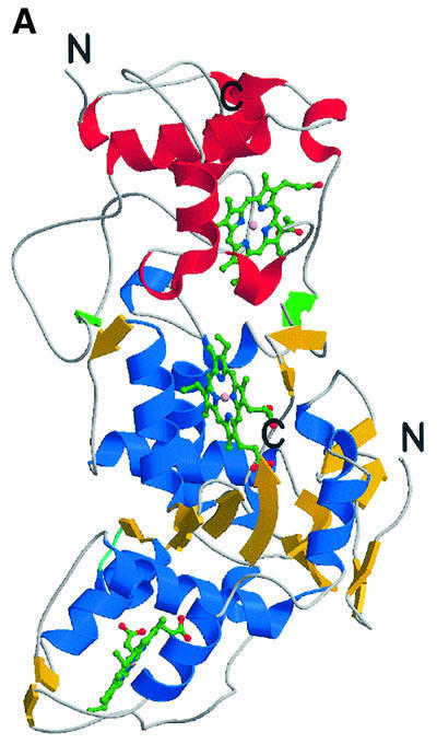
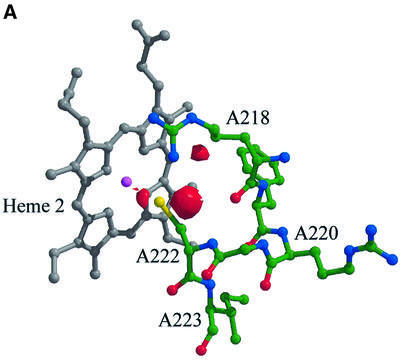
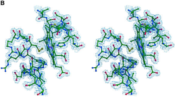
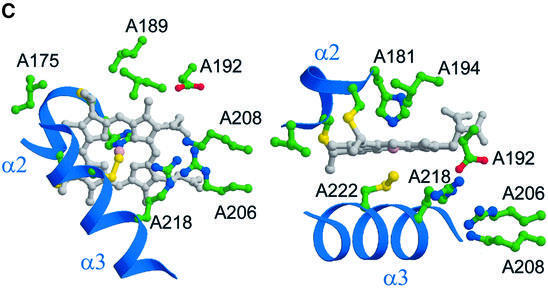
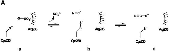
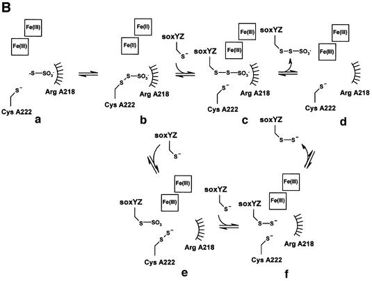
Reduced inorganic sulfur compounds are utilized by many bacteria as electron donors to photosynthetic or respiratory electron transport chains. This metabolism is a key component of the biogeochemical sulfur cycle. The SoxAX protein is a heterodimeric c-type cytochrome involved in thiosulfate oxidation. The crystal structures of SoxAX from the photosynthetic bacterium Rhodovulum sulfidophilum have been solved at 1.75 A resolution in the oxidized state and at 1.5 A resolution in the dithionite-reduced state, providing the first structural insights into the enzymatic oxidation of thiosulfate. The SoxAX active site contains a haem with unprecedented cysteine persulfide (cysteine sulfane) coordination. This unusual post-translational modification is also seen in sulfurtransferases such as rhodanese. Intriguingly, this enzyme shares further active site characteristics with SoxAX such as an adjacent conserved arginine residue and a strongly positive electrostatic potential. These similarities have allowed us to suggest a catalytic mechanism for enzymatic thiosulfate oxidation. The atomic coordinates and experimental structure factors have been deposited in the PDB with the accession codes 1H31, 1H32 and 1H33.


Figures

None
Fig. 1. The Friedrich–Kelly model for the Sox mechanism of thiosulfate oxidation in R.sulfidophilum. SoxAX catalyses a two-electron oxidation in which the sulfane sulfur of thiosulfate is linked to the antepenultimate cysteine residue of SoxY to produce SoxY–thiocysteine- S-sulfate. The figure is modified from Friedrich et al. (2001). Open circles represent sulfur atoms, whilst oxygen atoms are shown as filled circles.
None
Fig. 2. Amino acid sequences of SoxA and SoxX subunits from different sulfur-oxidizing bacteria. (A and B) Primary sequence alignments and haem coordination in the R.sulfidophilum SoxA and SoxX subunits, respectively. Sequences are those of the SoxAX proteins from R.sulfidophilum (Appia-Ayme et al., 2001) (Rs), Paracoccus denitrificans (Friedrich et al., 2000) (Pd), Chlorobium limicola (Klarskov et al., 1998) (Cl), Chlorobium tepidum (The Institute for Genomic Research. URL http://www.tigr.org) (Ct), Aquifex aeolicus (Deckert et al., 1998) (Aa) and Rhodopseudomonas palustris (DOE Joint Genome Institute. URL http://www.jgi.doe.gov/JGI_microbial/rhodo_homepage.html) (Rp). The alignments were generated with the program ClustalW using default parameters (see http://www.ebi.ac.uk/clustalw/). Secondary structural elements (β-sheet shown by blue arrows and α-helices by red bars) are indicated above the R.sulfidophilum sequence. The four residues indicated by Ptitsyn (1998) to be diagnostic for formation of each cytochrome c fold are highlighted in green, and the four conserved helices of the folds of the two domains (α1–α4 and α1′–α4′) are labelled. CXXCH haem-binding motifs and second axial ligands are indicated in yellow. Polypeptides contributing to the interface of the SoxAX complex and forming part of interface region A (see text) are boxed in blue; those forming part of interface region B are boxed in red. A consensus sequence showing identical residues is also given.
None
Fig. 3. The overall fold of the SoxAX complex. (A) The architecture of the complex. α-helices are shown in blue, and β-strands in yellow for SoxA, whilst for SoxX they are shown in red and green, respectively. The SoxA subunit is composed of two cytochrome c domains related by a pseudo 2-fold axis and contains two c-type haems with histidine–cysteine axial ligands. SoxX consists of a single cytochrome c fold with a histidine– methionine co-ordinated haem. (B) A stereo view of the Cα trace of the SoxAX molecule with every twentieth residue labelled. The SoxX subunit is coloured yellow, whilst for SoxA, the N-terminal 50 amino acids are coloured grey and the SoxAN and SoxAC cytochrome c domains are coloured blue and red, respectively. An insertion in the SoxX sequence from R.sulfidophilum relative to other cytochromes c (shown in purple) forms a major component of the subunit interface. (C) A close-up view of the electrostatic potential at the solvent-accessible molecular surface showing the substrate access channel and the binding groove for the C-terminal peptide of SoxYZ. The positions of arginine, lysine and histidine residues contributing to a basic substrate access funnel are shown. (D) Comparison of SoxA (left) with the two-domain cytochrome c fold of cytochrome c4 from Pseudomonas stutzeri (right). The C-terminal domains of SoxA and cytochrome c4 (yellow haems) are presented in the same orientation as determined by DALI (Holm and Sander, 1993). The four conserved helices (α1–α4) of the cytochrome c fold are shown for each domain in red. The N-terminal 50 amino acids of SoxA (shown in grey) pack against the face of the SoxAC domain, which in the previously characterized two-domain cytochromes c interacts with the N-terminal domains.
None
Fig. 4. The active site of SoxAX. (A and B) Post-translational modification of the haem 2 axial ligand. (A) The (Fo – Fc) Fourier map contoured at the 3.5σ level (shown in red), where σ represents the r.m.s. electron density for the unit cell. This map has been calculated using structure factor amplitudes and phases calculated from a refined model with a cysteine at residue A222. Note the small region of difference electron density adjacent to the side chain of ArgA218. This feature is also found in (Fo – Fc) maps calculated using the final refined structural model containing a cysteine persulfide haem ligand. The atoms and chemical bonds of haem 2 are coloured grey, except the haem iron which is magenta. (B) Stereo view of the (2Fo – Fc) electron density map around haem 2 calculated at 1.75 Å resolution and contoured at the 1.2σ level. This map has been calculated from a refined structural model with a cysteine persulfide at residue A222. (C) Orthogonal views of the active site region. CysA222 and HisA181 provide the axial ligands to the iron atom of haem 2. The basic residues A218, A206 and A208 form a possible proton-transfer relay from the active site.
None
Fig. 5. Insights into the mechanism of thiosulfate oxidation. (A) The mechanism of the sulfurtransferase, rhodanese. (B) The proposed mechanisms for thiosulfate oxidation by SoxAX. Note that the point at which reoxidation of haems 2 and 3 occurs is not known.
None
Fig. 5. Insights into the mechanism of thiosulfate oxidation. (A) The mechanism of the sulfurtransferase, rhodanese. (B) The proposed mechanisms for thiosulfate oxidation by SoxAX. Note that the point at which reoxidation of haems 2 and 3 occurs is not known.

Similar articles

Cited by

References

    1. Appia-Ayme C., Little,P.J., Matsumoto,Y., Leech,A.P. and Berks,B.C. (2001) A cytochrome complex essential for photosynthetic oxidation of both thiosulfate and sulfide in Rhodovulum sulfidophilum. J. Bacteriol., 183, 6107–6118. - PMC - PubMed
    1. Bartsch R.G. (1971) Cytochromes: bacterial. Methods Enzymol., 23, 344–363.
    1. Beinert H. (1983) Semi-micro methods for analysis of labile sulfide and of labile sulfide plus sulfane sulfur in unusually stable iron–sulfur proteins. Anal. Biochem., 131, 373–378. - PubMed
    1. Bordo D., Deriu,D., Colnaghi,R., Carpen,A., Pagani,S. and Bolognesi,M. (2000) The crystal structure of a sulfurtransferase from Azotobacter vinelandii highlights the evolutionary relationship between the rhodanese and phosphatase enzyme families. J. Mol. Biol., 298, 691–704. - PubMed
    1. Brune D.C. (1989) Sulfur oxidation by phototrophic bacteria. Biochim. Biophys. Acta, 975, 189–221. - PubMed

Publication types