. 2002 Nov 1;21(21):5682-90.
doi: 10.1093/emboj/cdf573.

The behaviour of polyamino acids reveals an inverse side chain effect in amyloid structure formation

Affiliations

The behaviour of polyamino acids reveals an inverse side chain effect in amyloid structure formation

Marcus Fändrich et al. EMBO J. .

Abstract

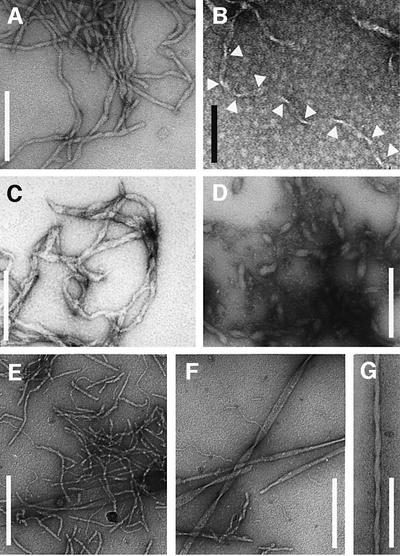
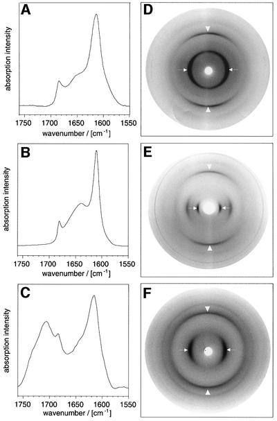
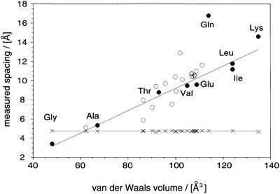
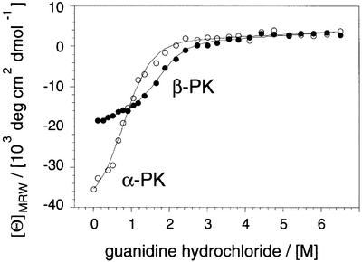
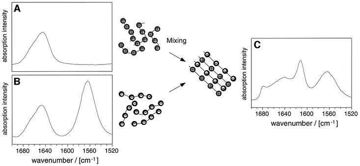
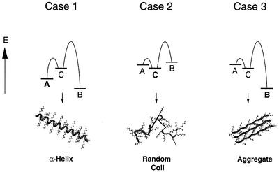
Amyloid fibrils and prions are proteinaceous aggregates that are based on a unique form of polypeptide configuration, termed cross-beta structure. Using a group of chemically distinct polyamino acids, we show here that the existence of such a structure does not require the presence of specific side chain interactions or sequence patterns. These observations firmly establish that amyloid formation and protein folding represent two fundamentally different ways of organizing polypeptides into ordered conformations. Protein folding depends critically on the presence of distinctive side chain sequences and produces a unique globular fold. By contrast, the properties of different polyamino acids suggest that amyloid formation arises primarily from main chain interactions that are, in some environments, overruled by specific side chain contacts. This side chain effect can be thought of as the inverse of the one that characterizes protein folding. Conditions including Alzheimer's and Creutzfeldt-Jakob diseases represent, on this basis, pathological cases in which a natural polypeptide chain has aberrantly adopted the conformation that is primarily defined by main chain interactions and not the structure that is determined by specific side chain contacts that depend on the polypeptide sequence.


Figures

None
Fig. 1. Far-UV CD spectra of different conformations of poly-l-lysine. The far-UV CD spectra of 0.1 mg/ml poly-l-lysine in an α-helical (filled circles, freshly dissolved PK at pH 11.1), β-sheet (open circles, PK at pH 11.1 after heating for 15 min, 52°C) and random coil conformation (diamonds, freshly dissolved PK in pure water).
None
Fig. 2. Morphology of PAA fibrils defined by EM. PK: (A) 2.5 mg/ml, H2O, pH 11.2, 65°C, 4 days or (B) 1 mg/ml, H2O, pH 10.1, 65°C, 1 week. Arrowheads emphasize the constrictions of a ribbon-like morphology. PE: (C) 1 mg/ml, D2O, pD 4.08, 65°C, 2 days or (D) H2O, pH 4.1, 65°C, 6 days. PT: 10 mg/ml (E and F) or 1 mg/ml (G) PT in 50 mM sodium borate pH 9.0 was exposed for 4 days to 65°C. Samples in (E) and (F) subsequently were kept at room temperature for 6 weeks. Scale bars: 200 nm (white), 100 nm (black).
None
Fig. 3. Internal conformations of fibrillar PAAs. FTIR amide I′ region of PT (A), PK (B) and PE (C) in D2O. Maxima: 1614 and 1685/cm (A); 1611 and 1680/cm (B); and 1616, 1683 and 1706/cm (C). X-ray diffraction pictures of films cast from PT (D), PK (E) and PE (F). To obtain the β-form of PE, the PAA was dissolved at 25 mg/ml in D2O, pD 4.1 and heated for 4 h to 95°C. Arrowheads are parallel to the plane of the film and show the main chain spacing; the arrows point to the side chain spacing.
None
Fig. 4. Comparison of the internal structure of PAA fibrils and other amyloid structures. Correlation of the main chain spacing (crosses) and the side chain spacing (circles) with the residual volume (Creighton, 1993). Closed symbols, PAAs (Arnott et al., 1967; Komoto et al., 1974; Lotz, 1974; Perutz et al., 1994); open symbols, heterogeneous sequences (Glenner et al., 1974; Inouye et al., 1993, 2000; Nguyen et al., 1995; Weaver, et al., 1996; Symmons et al., 1997; Kirschner et al., 1998; Malinchik et al., 1998; Groß et al., 1999; Luckey et al., 2000; Serpell et al., 2000). A linear fit to the PAA side chain spacings (excluding glutamine) is shown.
None
Fig. 5. Stability of α-PK and β-PK against denaturation. Guanidine hydrochloride denaturation of α-PK (222 nm, open symbols) and β-PK (217 nm, closed symbols) monitored by CD and fitted to a two-state transition (Santoro and Bolen, 1988).
None
Fig. 6. Formation of mixed aggregates from PE and PK. PK (A) and PE (B) do not aggregate in D2O, pD 7.5. The FTIR spectra show a random coil structure. On mixing the two solutions, the PE and PK aggregates are formed (C). FTIR spectra were recorded in D2O, pD 7.5. The total free amino acid concentrations were all 0.1 M. Amide I′ maxima: 1646 cm–1 (PE and PK in isolation); 1612, 1679 and 1642 cm–1 (mixed aggregates). The carboxylate group gives rise to a peak at 1565 cm–1.
None
Fig. 7. The partitioning between folding and amyloid formation. Polypeptides that can adopt ordered states with low contact order (A or α-PK), an ordered state with high contact order (B or β-PK) and a random coil state (C) show different relative energies under different physico-chemical conditions (cases 1–3). For example, the PK side chains have properties that destabilize both states A and B at neutral pH (case 2). Upon increasing the pH, unfavourable electrostatic effects are reduced. Since A is kinetically favourable over B (case 1), the latter state can only be observed under conditions that destabilize A, such as mild heating (case 3).

References

    1. Abkevich V.I., Gutin,A.M. and Shakhnovich,E.I. (1998) Theory of kinetic partitioning in protein folding with possible applications to prions. Proteins Struct. Funct. Genet., 31, 335–344. - PubMed
    1. Anfinsen C.B. (1973) Principles that govern the folding of protein chains. Science, 181, 223–230. - PubMed
    1. Arnott S., Dover,S.D. and Elliott,A. (1967) Structure of β-poly-l-alanine: refined atomic co-ordinates for an anti-parallel β-pleated sheet. J. Mol. Biol., 30, 201–208. - PubMed
    1. Avbelj F. and Baldwin,R.L. (2002) Role of backbone solvation in determining thermodynamic β propensities of the amino acids. Proc. Natl Acad. Sci. USA, 99, 1309–1313. - PMC - PubMed
    1. Baker D. (2000) A surprising simplicity to protein folding. Nature, 405, 39–42. - PubMed

Publication types