. 2002 Nov 1;21(21):5691-700.
doi: 10.1093/emboj/cdf568.

The cytoplasmic domain of the LDL receptor-related protein regulates multiple steps in APP processing

Affiliations

The cytoplasmic domain of the LDL receptor-related protein regulates multiple steps in APP processing

Claus U Pietrzik et al. EMBO J. .

Abstract

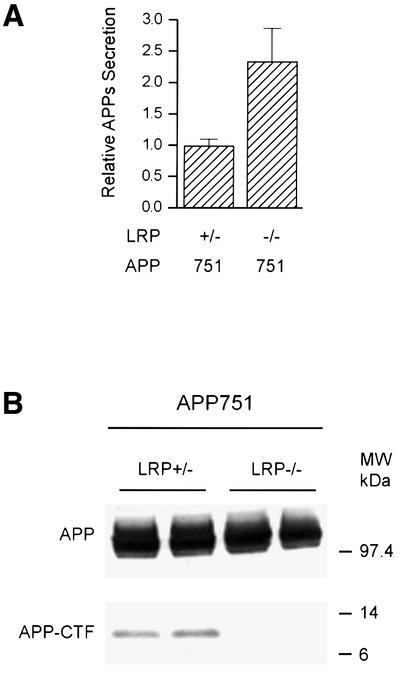
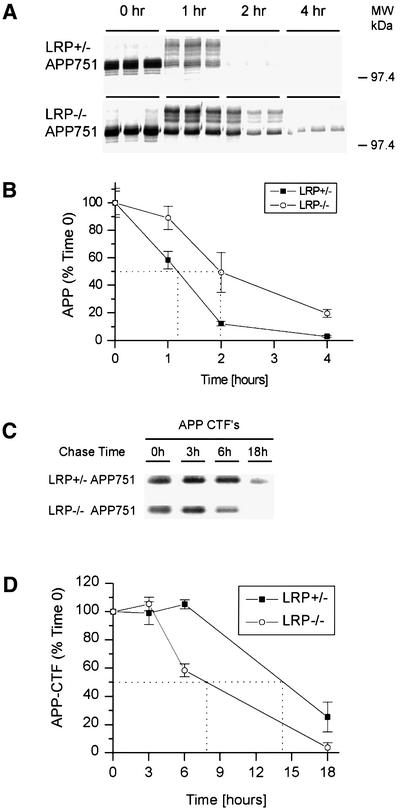
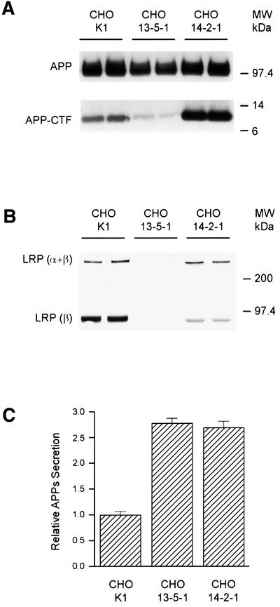
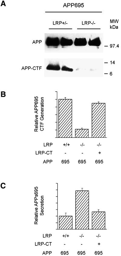
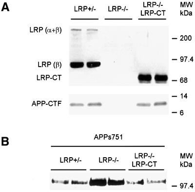
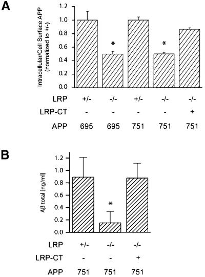
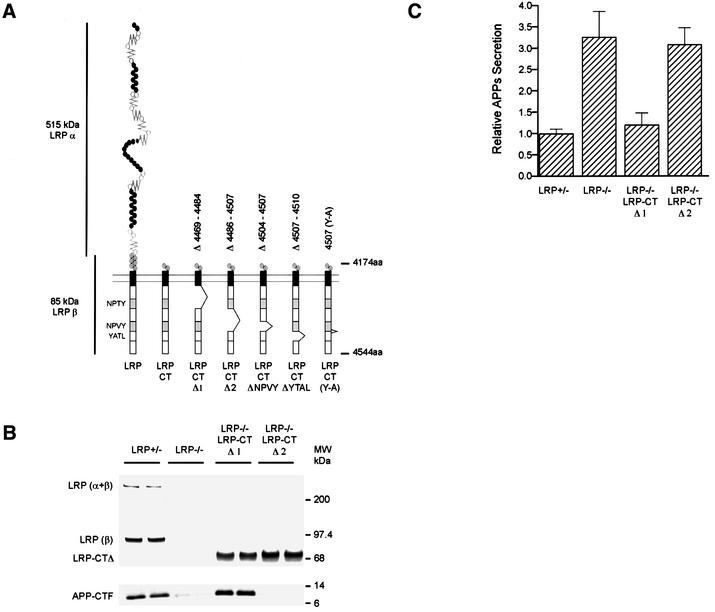
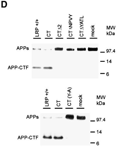
The low-density lipoprotein receptor-related protein (LRP) has recently been implicated in numerous intracellular signaling functions, as well as in Alzheimer's disease pathogenesis. Studies have shown that the beta-amyloid precursor protein (APP) interacts with LRP and that this association may impact the production of amyloid beta-protein (Abeta). In this report, we provide evidence that LRP regulates trafficking of intracellular proteins independently of its lipoprotein receptor functions. We show that in the absence of LRP, Abeta production, APP secretion, APP internalization, turnover of full-length APP and stability of APP C-terminal fragments are affected. Importantly, these changes are not APP isoform dependent. Using deletion constructs, the critical region in LRP that modulates APP processing was mapped to a seven peptide domain around the second NPXY domain (residues 4504-4510). Therefore, we propose a model by which LRP functionally modulates APP processing, including those steps critical for Abeta production, through interactions of the cytosolic domains.


Figures

None
Fig. 1. APPs and APP-CTF levels in LRP–/– cells. Mouse embryonic fibroblasts lacking the LRP gene (LRP–/–) and corresponding LRP-expressing control fibroblasts (LRP+/–) were stably transfected with APP751. APPs was immunoprecipitated using the monoclonal antibody 1G7-5A3 and the samples immunoblotted with an APP polyclonal antibody (863) as described in Materials and methods. (A) LRP-deficient cells secrete ∼2.5-fold more APPs than LRP-expressing cells. Results are expressed as the average of two experiments in triplicate ± SEM. (B) APP expression in LRP+/– and LRP–/– fibroblasts was determined by immunoblotting with a polyclonal antibody (CT15) raised against the C-terminus of APP. Single clones of similar APP expression were selected (top panel). In LRP–/– cells, there is a dramatic reduction in the level of APP-αCTF (bottom panel).
None
Fig. 2. Turnover of full-length APP and APP-αCTF in LRP–/– cells. LRP+/– and LRP–/– fibroblasts were pulse labeled with [35S]methionine/cysteine for 15 min and chased for 0, 1, 2 and 4 h (A). At time 0, APP consists predominantly of immature N-glycosylated species. Both the N-glycosylated and mature N- and O-glycosylated species are abundant at 1 h for both cell types. After a 2 h chase period, the APP level is dramatically reduced in LRP+/– cells and hardly detectable at 4 h. In contrast, APP can still be detected in LRP–/– cells even after a 4 h chase period. (B) The half-life was determined by quantitating the results from (A) as shown. (C) APP-αCTF turnover was determined by metabolically labeling LRP+/– and LRP–/– fibroblasts with [35S]methionine/cysteine for 1 h and chasing for 3, 6 and 18 h. Similar amounts of APP-αCTFs are present after the labeling and after the 3 h chase period in LRP+/– and LRP–/– cells. Even after an 18 h chase period, APP-αCTFs can still be detected in LRP+/– fibroblasts. However, APP-αCTFs in LRP–/– fibroblasts were almost completely degraded. (D) The half-life of APP-αCTF was determined by quantitating the results from (C) as shown. The experiment in (A) was performed in triplicate and that in (C) in duplicate, and representative experiments are shown.
None
Fig. 3. APP processing in CHO cell lines. (A) Endogenous full-length APP (upper panel) and APP-CTF (lower panel) were immunoblotted with APP C-terminal antibody (CT15) from the CHO cell line 13-5-1 deficient in LRP, the CHO cell line 14-2-1 expressing a mutant LRP defective in trafficking to the plasma membrane, and the control CHO K1 cell line. Note that cells lacking LRP (13-5-1) show a significant reduction in the level of APP-αCTF (bottom panel), while the cells expressing the LRP trafficking mutant (14-2-1) show higher endogenous APP-αCTF levels. (B) LRP is detected by immunoblotting with the polyclonal antibody 1704 raised against the C-terminus of LRP. As expected, no LRP can be seen in the CHO cell line 13-5-1 (middle lanes), while the amount of β-subunit in CHO 14-2-1 cells is significantly less than in control cells, with the ratio indicating lack of furin cleavage due to retention in the early secretory compartments. Uncleaved full-length LRP (α + β) can be visualized and the levels are comparable between CHO 14-2-1 and CHO K1 control cells. (C) Secretion of endogenous APPs into medium was increased in both CHO 13-5-1 and CHO 14-2-1 cells as compared with control CHO K1 cells.
None
Fig. 4. Alterations of APP processing in LRP–/– cells are isoform independent. (A) LRP+/– and LRP–/– cells stably transfected with APP695 were analyzed for levels of APP-αCTFs. Single clones of similar APP expression were analyzed (top panel). Note the dramatic reduction in APP-CTF expression in LRP –/– cells (bottom panel). Quantitation of APP-αCTF (B) and APPs (C) levels in LRP-deficient CHO 13-5-1 and control CHO cells transfected with APP695. Note that APP-αCTF and APPs derived from APP695 were altered similarly in LRP-deficient cells as those from APP751. Introduction of LRP-CT in the LRP- deficient cells restored the abnormalities in APP-αCTF (B) and APPs (C) back to control levels. Results are the average ± SEM (n = 2).
None
Fig. 5. Processing of APP751 in LRP–/– cells can be restored by expression of an LRP-CT construct. (A) LRP–/– fibroblasts transfected with APP751 were infected with a retrovirus expressing the LRP-CT construct and immunoblotted with the LRP C-terminal antibody 1704. No signal is seen in the LRP–/– cells (middle lanes, top panel). The LRP-CT protein is a truncated β-subunit (right lanes, LRP–/– LRP-CT) and migrates faster than the authentic β-subunit (left lanes, LRP+/–) and, as expected, the full-length LRP species (α + β) is absent in the LRP–/– LRP-CT cells (right lanes, top panel). In the bottom panel, immunoblotting of APP-αCTF was carried out with the APP C-terminal antibody CT15. Note that following expression of the LRP-CT construct in LRP–/– cells, the normally low levels of APP-CTF in LRP–/– cells (middle lanes, bottom panel) are now restored to the level seen in LRP+/– cells (compare right with left lanes, bottom panel). (B) Levels of APPs in medium of LRP+/–, LRP–/– and LRP–/– LRP-CT cells were determined by immunoprecipitation/western blotting as before. As with APP-CTF levels, expression of LRP-CT in LRP–/– cells restores the abnormal levels of APPs. Similarly to APP695, APPs release derived from APP751 is decreased after introduction of LRP-CT in LRP–/– cells.
None
Fig. 6. APP internalization and Aβ secretion in LRP–/– cells. (A) Steady-state internalization of APP from the cell surface was measured with iodinated 1G7 antibody at 37°C (see Materials and methods). In LRP+/– control cells expressing either APP695 or APP751, ∼55% of APP is internalized. In this experiment, the ratios of internalized to cell surface APP are normalized to the LRP+/– control cells. LRP–/– cells stably transfected with APP751 or APP695 show a 50% reduction in APP endocytosis as compared with control (LRP+/–) cells. The reduction in APP internalization in LRP–/– cells is restored similarly after expression of the LRP-CT construct. (B) Reduction in Aβ release in LRP–/– cells is rescued by expression of the LRP-CT construct. Aβ was measured by ELISA from LRP–/–, LRP–/–LRP-CT and LRP+/– cells after 72 h collection. The Aβ level is reduced 5-fold in LRP–/– cells transfected with APP751 as compared with control LRP+/– cells transfected with APP751, normalized for the level of APP expression. The reduction in Aβ in LRP–/– cells is restored to control level after expression of the LRP-CT construct. All panels show the results (average ± SEM) of representative experiments performed in triplicate. Statistical analysis was performed using Student’s t-test (*P < 0.05).
None
Fig. 7. The region around the second NPXY domain in LRP regulates APP processing. (A) Schematic illustration of two LRP-CT deletion constructs, LRP-CTΔ1 (lacking amino acids 4469–4484) or LRP-CTΔ2 (lacking amino acids 4486–4507). (B) Expression of LRP-CTΔ1 and LRP-CTΔ2 in LRP–/– cells is comparable to the endogenous level of LRP as seen by immunoblotting with LRP antibody 1704 (upper panel). The reduction in the levels of APP-CTFs in LRP–/– cells is restored by expression of the LRP-CTΔ1 but not the LRP-CTΔ2 construct. (C) Consistent with the results shown in (B), APPs secretion was restored to control LRP+/– levels only in the LRP-CTΔ1-transfected cell line but not in the LRP-CTΔ2 cell line. (D) LRP-CTΔNPVY (lacking amino acids 4504–4507), LRP-CTΔYATL (lacking amino acids 4507–4510) and LRP-CT (Y4507A) were expressed in LRP-deficient CHO 13-5-1 cells together with APP695 [see (A) for schematic]. Similarly to LRP-CTΔ2, expression of neither LRP-CTΔNPVY (top panel), LRP-CTΔYATL (top panel) nor LRP-CT (Y4507A) (bottom panel) was able to restore the reduction in APP-CTF levels or the increase in APPs to that seen in control K1 cells. In contrast, the expression of LRP-CT restored the deficiencies in APP processing in LRP–/– CHO 13-5-1 cells to control levels.

References

    1. Annaert W.G. et al. (1999) Presenilin 1 controls γ-secretase processing of amyloid precursor protein in pre-Golgi compartments of hippocampal neurons. J. Cell Biol., 147, 277–294. - PMC - PubMed
    1. Boucher P., Liu,P., Gotthardt,M. Hiesberger,T., Anderson,R.G. and Herz,J. (2002) Platelet-derived growth factor mediates tyrosine phosphorylation of the cytoplasmic domain of the low density lipoprotein receptor-related protein in caveolae. J. Biol. Chem., 277, 15507–15513. - PubMed
    1. Bu G. and Rennke,S. (1996) Receptor-associated protein is a folding chaperone for low density lipoprotein receptor-related protein. J. Biol. Chem., 271, 22218–22224. - PubMed
    1. Cao X. and Sudhof,T.C. (2001) A transcriptively active complex of APP with Fe65 and histone acetyltransferase Tip60. Science, 293, 115–120. - PubMed
    1. Cook D.G., Forman,M.S., Sung,J.C., Leight,S., Kolson,D.L., Iwatsubo,T., Lee,V.M. and Doms,R.W. (1997) Alzheimer’s A β(1–42) is generated in the endoplasmic reticulum/intermediate compartment of NT2N cells. Nat. Med., 3, 1021–1023. - PubMed

Publication types

MeSH terms

Substances