. 2002 Nov 1;21(21):5701-10.
doi: 10.1093/emboj/cdf559.

Reversible stages of the low-pH-triggered conformational change in influenza virus hemagglutinin

Affiliations

Reversible stages of the low-pH-triggered conformational change in influenza virus hemagglutinin

Eugenia Leikina et al. EMBO J. .

Abstract

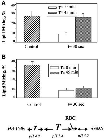
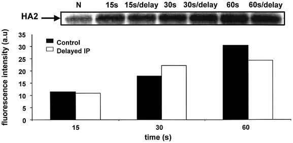
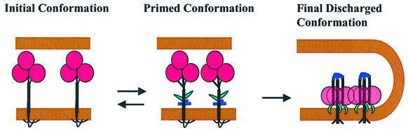
The refolding of the prototypic fusogenic protein hemagglutinin (HA) at the pH of fusion is considered to be a concerted and irreversible discharge of a loaded spring, with no distinct intermediates between the initial and final conformations. Here, we show that HA refolding involves reversible conformations with a lifetime of minutes. After reneutralization, low pH-activated HA returns from the conformations wherein both the fusion peptide and the kinked loop of the HA2 subunit are exposed, but the HA1 subunits have not yet dissociated, to a structure indistinguishable from the initial one in functional, biochemical and immunological characteristics. The rate of the transition from reversible conformations to irreversible refolding depends on the pH and on the presence of target membrane. Importantly, recovery of the initial conformation is blocked by the interactions between adjacent HA trimers. The existence of the identified reversible stage of refolding can be crucial for allowing multiple copies of HA to synchronize their release of conformational energy, as required for fusion.


Figures

None
Fig. 1. Reversible conformations of low-pH-activated HA identified by means of a fusion inactivation assay. X31 (A) or Japan HA-cells (B) at 22°C were first treated with a 5 min pH 4.9 pulse (‘AP’) in the absence of target membrane. Then, the cells were incubated at neutral pH for different times. Finally, the cells were incubated with RBCs for 15 min and, after removal of unbound RBCs, the second (‘fusion-triggering’) low-pH pulse, FTP [a 2 min pH 4.9 pulse in (A) and a 5 min pH 5.2 pulse in (B)] was applied. Here and in the experiments reported in the following figures, the final extents of lipid mixing were measured by fluorescence microscopy. The total time intervals between the activating and fusion-triggering pulses were 15 min (bars 2) and 45 min (bars 3–7). Bars 4 and 5, thermolysin (25 µg/ml), and bars 6 and 7, DTT (10 mM) were applied for either the first (bars 4 and 6, respectively) or the last (bars 5 and 7, respectively) 5 min of incubation between the activating and fusion-triggering pulses. Fusion extents were normalized to those in the control experiments, in which the FTP [a 2 min pH 4.9 pulse in (A) and a 5 min pH 5.2 pulse in (B)] was applied to the HA-cells with bound RBCs untreated with an AP. Fusion extents of 83.9 and 63.5% were taken as 100% in bars A1 and B1, respectively.
None
Fig. 2. Changes in the accessibility of different HA epitopes at different stages of HA activation and recovery. X31 HA-cells were incubated at pH 4.9 for different times. (AD) HA conformation was analyzed by immunoprecipitation with different primary antibodies. LC89, antiserum against fusion peptide (FP), interfacial antibody (IF) or HC67 antibody (A–D, respectively) was applied following the low-pH pulse, either after an additional 30 min incubation at pH 7.4 or immediately after the pulse (marked in the pH 7.4 line under the panel as ‘30 ’or ‘–’, respectively). The notations ‘–’ and ‘+’, for pH 4.9 and 7.4, respectively, under the panels mark the control experiments, in which HA-cells were not treated with a low-pH pulse. (E) X31 HA-cells were treated with a 5 min pH 4.9 pulse at 22°C. Accessibility of the kinked loop epitope was assayed by CELISA, with LC89 antibodies applied immediately after the pulse (taken as 100%) or after an additional 10, 20, 30 or 45 min incubation at neutral pH following the pulse. (F) X31 HA-cells were incubated at pH 4.9 (28°C) for different times. Binding of LC89 (circles) and IF (triangles) antibodies was assayed by CELISA either immediately after the low-pH pulse (open symbols) or after an additional 15 min incubation at neutral pH following the pulse (closed symbols). After subtraction of non-specific binding, the results were normalized to the levels of the binding of the corresponding antibody, LC89 and IF, detected immediately after the 5 min pH 4.9 pulse.
None
Fig. 3. Recovery of low-pH-activated HA molecules at 37 and 4°C evaluated by fusion inactivation assay. (A) Rapid loss of reversibility at 37°C. X31 HA-cells at 37°C were first treated with an AP (pH 4.9 for 1, 2, 5 or 10 min) applied in the absence of target membrane. The cells were incubated at pH 7.4 for 0 or 30 min, incubated with RBCs for 15 min and treated with an FTP (1 min, pH 4.9). The time interval between the end of the AP and the beginning of the FTP was either 15 or 45 min. The ‘control’ bar presents the results of the control experiment, in which only the FTP was applied to the HA-cells, with bound RBCs untreated with an AP. The final extents of lipid mixing were measured by fluorescence microscopy. (B) HA recovery is blocked at 4°C. X31 HA-cells at 22°C were first treated with an AP (pH 4.9 for 5 or 10 min) applied in the absence of target membrane. The cells were incubated at pH 7.4 for 0 or 30 min, incubated with RBCs for 15 min and treated with an FTP (2 min, pH 4.9, 22°C). During the total time interval, either 15 or 45 min, between the end of the AP and the beginning of the FTP, cells were kept at either 22 or 4°C. In the experiment represented in the ‘control’ bar, the FTP (2 min, pH 4.9, 22°C) was applied to the HA-cells, with bound RBCs untreated with an AP.
None
Fig. 4. Inhibition of the recovery of low-pH-activated HA molecules in the presence of target membrane. X31 HA-cells at 22°C were first treated with an AP (1 min at pH 4.9) applied in the absence of target membrane. The cells were then incubated at pH 7.4 for 15 or 30 min, incubated with RBCs for 15 min and treated with an FTP (2 min pH 4.9). Alternatively, RBCs were added immediately after the end of the AP and were present throughout a 15, 30 or 45 min incubation between the activating and the fusion-triggering pulses. In the experiment shown in the ‘control’ bar, the FTP (2 min at pH 4.9) was applied to the HA-cells, with bound RBCs untreated with an AP.
None
Fig. 5. Effects of HA surface density on the recovery of the initial HA conformation. Japan HA-cells pre-incubated without (A) or with 5 mM NaBut (B) were first treated with an AP (30 s at pH 4.9, 22°C) in the absence of RBCs. Cells were then incubated at pH 7.4 for 0 or 30 min, incubated with RBCs for 15 min and treated with an FTP (2 min at pH 5.2). The time interval T between the end of the AP and the beginning of the FTP was either 15 or 45 min. In the experiment shown in the ‘control’ bar, the FTP (2 min at pH 5.2) was applied to the HA-cells, with bound RBCs untreated with an AP.
None
Fig. 6. Irreversibility of a low-pH-activated HA conformation with an exposed FP at influenza virus particles. Virus was treated with pH 4.9 at 22°C for 0 (marked as N), 15, 30 or 60 s. FP exposure was assayed by immunoprecipitation. The antiserum was added either immediately after reneutralization or after an additional 60 min incubation at pH 7.4. The intensity of the HA2 band in the presented gel was quantified and plotted in the bar chart, with the subtracted intensity of the band observed in the experiment without low-pH application. The total protein concentration loaded, determined by volume, is comparable in each lane, since we started the experiment with the same amount of virus for each sample.
None
Fig. 7. Schematic diagram depicting refolding of acidified HA trimers in the proposed pathway from the initial neutral-pH form to a primed reversible form, followed by the irreversible transition to a final low-pH form. For the sake of simplicity, the receptors in the target membrane are not shown. The structure of the primed form is shown here as characterized at the neutral pH, and might be somewhat different at low pH. The membranes, HA1, the exposed FP and the kinked loop of HA2 are shown in brown, pink, green and blue, respectively. Exposure of the FP and kinked loop is reversible. In contrast, the dissociation of the HA1 trimer occurs already at the irreversible stage of HA refolding. The transition from primed to final form is promoted by interactions between adjacent trimers.

Similar articles

Cited by

References

    1. Bullough P.A., Hughson,F.M., Skehel,J.J. and Wiley,D.C. (1994) Structure of influenza haemagglutinin at the pH of membrane fusion. Nature, 371, 37–43. - PubMed
    1. Carr C.M. and Kim,P.S. (1993) A spring-loaded mechanism for the conformational change of influenza hemagglutinin. Cell, 73, 823–832. - PubMed
    1. Chen J., Lee,K.H., Steinhauer,D.A., Stevens,D.J., Skehel,J.J. and Wiley,D.C. (1998) Structure of the hemagglutinin precursor cleavage site, a determinant of influenza pathogenicity and the origin of the labile conformation. Cell, 95, 409–417. - PubMed
    1. Chen J., Skehel,J.J. and Wiley,D.C. (1999) N- and C-terminal residues combine in the fusion-pH influenza hemagglutinin HA(2) subunit to form an N cap that terminates the triple-stranded coiled coil. Proc. Natl Acad. Sci. USA, 96, 8967–8972. - PMC - PubMed
    1. Chernomordik L.V., Leikina,E., Frolov,V., Bronk,P. and Zimmerberg,J. (1997) An early stage of membrane fusion mediated by the low pH conformation of influenza hemagglutinin depends upon membrane lipids. J. Cell Biol., 136, 81–94. - PMC - PubMed

Substances