. 2002 Nov 1;21(21):5733-44.
doi: 10.1093/emboj/cdf575.

Hinge-mediated dimerization of SMC protein is essential for its dynamic interaction with DNA

Affiliations

Hinge-mediated dimerization of SMC protein is essential for its dynamic interaction with DNA

Michiko Hirano et al. EMBO J. .

Abstract

Structural maintenance of chromosomes (SMC) proteins play central roles in regulating higher order chromosome dynamics from bacteria to humans. As judged by electron microscopy, the SMC homodimer from Bacillus subtilis (BsSMC) is composed of two antiparallel, coiled-coil arms with a flexible hinge. Site-directed cross-linking experiments show here that dimerization of BsSMC is mediated by a hinge-hinge interaction between self-folded monomers. This architecture is conserved in the eukaryotic SMC2-SMC4 heterodimer. Analysis of different deletion mutants of BsSMC unexpectedly reveals that the major DNA-binding activity does not reside in the catalytic ATPase domains located at the ends of a dimer. Instead, point mutations in the hinge domain that disturb dimerization of BsSMC drastically reduce its ability to interact with DNA. Proper hinge function is essential for BsSMC to recognize distinct DNA topology, and mutant proteins with altered hinge angles cross-link double-stranded DNA in a nucleotide-dependent manner. We propose that the hinge domain of SMC proteins is not a simple dimerization site, but rather it acts as an essential determinant of dynamic SMC-DNA interactions.


Figures

None
Fig. 1. Design and characterization of BsSMC mutants. (A) Wild-type BsSMC (GGGG) and its mutant derivatives (AAAA and hinge-less) used in a previous study (Hirano et al., 2001). In this diagram, it is postulated that dimerization of BsSMC is mediated by a hinge–hinge interaction (shown by ?). (B) The wild-type and mutant BsSMC proteins were purified, fractionated by SDS–PAGE and stained with Coomassie Blue. (C) The purified BsSMC proteins were fractionated by centrifugation on 5–20% sucrose gradients. Fractions were resolved by SDS–PAGE and stained with Coomassie Blue. The positions of three protein standards [ovalbumin (3.7S), BSA (4.6S) and aldolase (7.3S)] are indicated. The predicted structure for each construct is shown on the right.
None
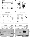
Fig. 2. The folding of BsSMC as revealed by site-directed, protein–protein cross-linking. (A) Two models for the folding of BsSMC. Dimerization may be mediated by coiled-coil interactions between two different subunits (model I). Alternatively, the two subunits may be self-folded to form two separate coiled-coil arms, which in turn dimerize by a hinge-mediated interaction (model II). (B) The positions of cysteine residues used in the cross-linking experiments. Two catalytic domains, each of which is composed of N- and C-terminal sequences, are shown. The open circles indicate the positions of the cysteine residues introduced into the N-terminal sequence (S55C and S142C). The filled stars indicate the positions of the naturally occurring cysteine (C1114) and the artificially introduced cysteine (C1070S) in the C-terminal sequence. Cross-linking is expected to occur between the two residues connected by the arches. (C and D) Predicted results from the site-directed cross-linking of the two-armed (GGGG) or single-armed (DDDD) protein. (E) Purified proteins were treated with BMH in the presence or absence of the indicated nucleotides (no, no nucleotide; T, 1 mM ATP; γS, 1 mM ATPγS), fractionated by 2.5–7.5% SDS–PAGE and analyzed by immunoblotting with an anti-BsSMC antibody. The no-cysteine mutant (lanes 1–3), single cysteine mutants (S142C, lanes 4–6; C1114, lanes 7–9) and the two-cysteine mutant (S142C-C1114, lanes 10–15) were used. The two-armed (GGGG; lanes 1–12) and single-armed (DDDD; lanes 13–15) versions were tested. Specific cross-linking products involving two cysteines at different positions (S142C and C1114) are shown by arrows. Background products involving cysteines at the same position (e.g. S142C of one polypeptide and S142C of the other) are shown by asterisks. These bands probably correspond to linear dimers, which are not depicted in (C) or (D). Non-cross-linked BsSMC polypeptides are shown by open triangles. (F) The same experiment was performed with a different set of mutants: the no-cysteine mutant (lanes 1–3); single-cysteine mutants (S55C, lanes 4–6; S1070C, lanes 7–9); and the two-cysteine mutant (S55C-S1070C, lanes 10–15). The two-armed (GGGG; lanes 1–12) and single-armed (DDDD; lanes 13–15) versions were tested.
None
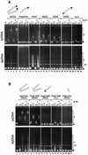
Fig. 3. DNA-binding activities of BsSMC mutants. (A) A fixed amount of ssDNA (top; 15.6 µM nucleotides) or negatively supercoiled dsDNA (bottom; 15.6 µM nucleotides) was incubated with two different concentrations of proteins (210 and 420 nM arms) in a buffer containing 5 mM KCl in the presence or absence of 1 mM ATP. No protein was added in lane 1. The reaction mixtures were fractionated on a 0.7% agarose gel and visualized by ethidium bromide stain. Protein–DNA complexes are indicated by asterisks, and free DNAs are indicated by arrows. NC indicates a nicked-circular population present in the dsDNA substrate. (B) The same assay was performed as above using a series of deletion mutants.
None
Fig. 4. ATPase activities of wild-type protein (GGGG), hinge mutants (AAAA, GGDD, AADD and DDDD) and the hinge-less mutant under different conditions. KCl titration at 2 mM MgCl2 (panels 1 and 2), MgCl2 titration at 5 mM KCl (panels 3 and 4) and MgCl2 titration at 50 mM KCl (panels 5 and 6) in the absence (panels 1, 3 and 5) or presence (panels 2, 4 and 6) of ssDNA (φX174 virion DNA; 31.2 µM nucleotides) are shown. A fixed protein concentration of 300 nM arms (equivalent to 150 nM dimers in the case of wild-type BsSMC) was used for all the proteins. The rate of ATP hydrolysis is expressed as the number of ATP molecules hydrolyzed per second per arm.
None
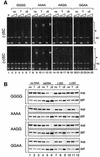
Fig. 5. Altered DNA-binding properties of hinge mutants. (A) Gel shift assay. A fixed amount of negatively supercoiled DNA (top; 15.6 µM nucleotides) or positively supercoiled DNA (bottom; 15.6 µM nucleotides) was incubated with two different concentrations of proteins (210 and 420 nM arms) in a buffer containing 5 mM KCl in the presence or absence of the indicated nucleotides (no, no nucleotide; T, 1 mM ATP; γS, 1 mM ATPγS). The reaction mixtures were fractionated on a 0.7% agarose gel and visualized by ethidium bromide stain. Protein–DNA complexes are indicated by asterisks. The positions of nicked circular DNA (NC), negative (–) and positive (+) supercoiled DNA are also shown. (B) Spin-down assay. A fixed amount of BsSMC proteins was mixed with either no DNA, ssDNA, negatively supercoiled DNA [(–)SC] or positively supercoiled DNA [(+)SC], in the presence or absence of the indicated nucleotides (no, no nucleotide; T, 1 mM ATP; γS, 1 mM ATPγS). After incubation, the mixtures were spun at 16 000 g for 15 min. The supernatants (sup) and pellets (ppt) were separated, fractionated by 7.5% SDS–PAGE and stained with silver.
None
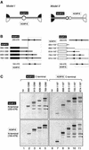
Fig. 6. The folding of the SMC2–SMC4 heterodimer as revealed by a coiled-coil interaction assay. (A) Two models for the folding of hCAP-C/SMC4 and hCAP-E/SMC2. Dimerization may be mediated by coiled-coil interactions between hCAP-C and hCAP-E (model I). Alternatively, the hCAP-C and hCAP-E subunits may be self-folded to form two separate coiled-coil arms, which in turn dimerize by a hinge-mediated interaction (model II). (B) Left: a deletion series of fragments containing the C-terminal domain of hCAP-C was co-translated in vitro with an N-terminal coiled-coil fragment of hCAP-C (253–576) or an N-terminal coiled-coil fragment of hCAP-E (164–479). Right: alternatively, a deletion series of fragments containing the C-terminal domain of hCAP-E was co-translated in vitro with one of the two N-terminal coiled-coil fragments. (C) The translation reactions were immunoprecipitated with antibodies that specifically recognize the C-terminal domain of hCAP-C (lanes 1–5) or hCAP-E (lanes 6–11). After washing, the immunoprecipitates were fractionated by SDS–PAGE and analyzed by autoradiography. Co-precipitated N-terminal fragments are indicated by the asterisks. In vitro translation of the N-terminal domain of hCAP-C or hCAP-E produced two bands, the smaller one of which is likely to be a translation product starting from an internal methionine. The combinations of fragments that interact with each other are boxed in the diagrams shown in (B).
None
Fig. 7. A hypothetical model for BsSMC–DNA interactions. Wild-type BsSMC (GGGG), which has a flexible hinge (shown by the white ovals), displays a variety of conformations and supports dynamic interactions with DNA. The AAAA mutant, which has a fixed and open hinge (shown by the black ovals), supports a robust dimer–dimer interaction when the catalytic domains of two different dimers associate with each other in the presence of ATPγS.

References

    1. Akhmedov A.T., Frei,C., Tsai-Pflugfelder,M., Kemper,B., Gasser,S.M. and Jessberger,R. (1998) Structural maintenance of chromosomes protein C-terminal domains bind preferentially to DNA with secondary structure. J. Biol. Chem., 273, 24088–24094. - PubMed
    1. Akhmedov A.T., Gross,B. and Jessberger,R. (1999) Mammalian SMC3 C-terminal and coiled-coil protein domains specifically bind palindromic DNA, do not block DNA ends and prevent DNA bending. J. Biol. Chem., 274, 38216–38224. - PubMed
    1. Anderson D.E., Losada,A., Erickson,H.P. and Hirano,T. (2002) Condensin and cohesin display different arm conformations with characteristic hinge angles. J. Cell Biol., 156, 419–424. - PMC - PubMed
    1. Bazett-Jones D.P., Kimura,K. and Hirano,T. (2002) Efficient supercoiling of DNA by a single condensin complex as revealed by electron spectroscopic imaging. Mol. Cell, 9, 1183–1190. - PubMed
    1. Britton R.A., Lin,D.C.-H. and Grossman,A.D. (1998) Characterization of a prokaryotic SMC protein involved in chromosome partitioning. Genes Dev., 12, 1254–1259. - PMC - PubMed

Publication types

MeSH terms