. 2002 Nov 1;21(21):5833-42.
doi: 10.1093/emboj/cdf569.

Importin alpha can migrate into the nucleus in an importin beta- and Ran-independent manner

Affiliations

Importin alpha can migrate into the nucleus in an importin beta- and Ran-independent manner

Yoichi Miyamoto et al. EMBO J. .

Abstract

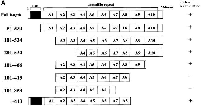
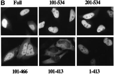
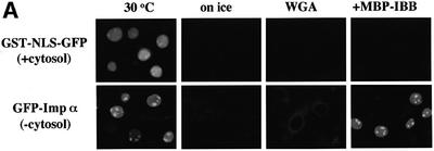
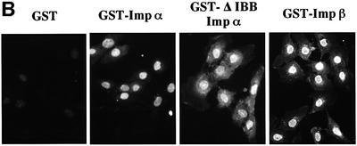
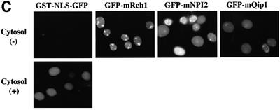
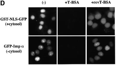
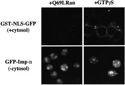
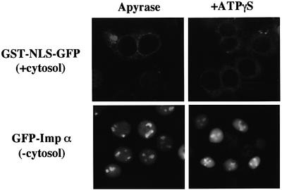
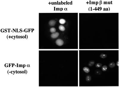
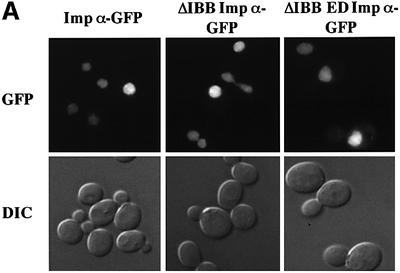
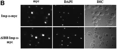
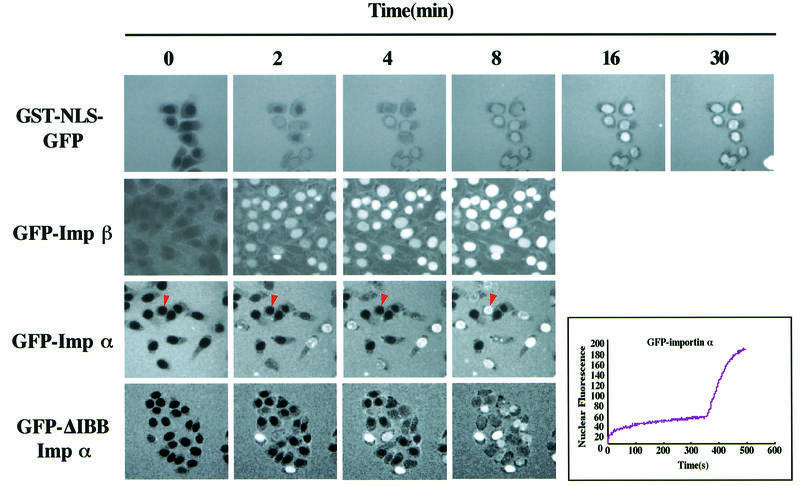
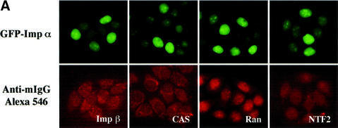
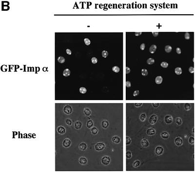
A classical nuclear localization signal (NLS)-containing protein is transported into the nucleus via the formation of a NLS-substrate/importin alpha/beta complex. In this study, we found that importin alpha migrated into the nucleus without the addition of importin beta, Ran or any other soluble factors in an in vitro transport assay. A mutant importin alpha lacking the importin beta-binding domain efficiently entered the nucleus. Competition experiments showed that this import pathway for importin alpha is distinct from that of importin beta. These results indicate that importin alpha alone can enter the nucleus via a novel pathway in an importin beta- and Ran-independent manner. Furthermore, this process is evolutionarily conserved as similar results were obtained in Saccharomyces cerevisiae. Moreover, the import rate of importin alpha differed among individual nuclei of permeabilized cells, as demonstrated by time-lapse experiments. This heterogeneous nuclear accumulation of importin alpha was affected by the addition of ATP, but not ATPgammaS. These results suggest that the nuclear import machinery for importin alpha at individual nuclear pore complexes may be regulated by reaction(s) that require ATP hydrolysis.


Figures

None
Fig. 1. Nuclear accumulation of importin α lacking an IBB domain. (A) Summary and the cellular localization of the importin α (mNPI2) deletion mutants used in this transfection analysis. These mutants were constructed into pEGFP-C2 transfection vectors (see Materials and methods). (B) Cellular localization of these mutants in transiently transfected HeLa cells. After transfection (12 h), the cells were fixed with 3.7% formaldehyde in PBS and the subcellular localization of EGFP-fused proteins was observed.
None
Fig. 2. Importin α is able to migrate into the nucleus in an importin β-independent manner in an in vitro assay. (A–D) Cells were treated with 40 µg/ml digitonin in TB (see Materials and methods) for 5 min on ice, and after washing with PBS twice, the cells were incubated with 10 µl of testing solution. (A) Digitonin-permeabilized MDBK cells were incubated with 2.5 µM GFP–importin α (mRch1) with TB alone or 2.5 µM GST–NLS–GFP with cytosolic extracts prepared from mouse Ehrlich ascites tumor cells and an ATP regeneration system for 20 min at 30°C or on ice. The other import reactions of GFP–importin α or GST–NLS–GFP were performed for 20 min at 30°C after pretreatment with 0.4 mg/ml WGA for 10 min at 30°C, or in the presence of 25 µM MBP–IBB for 20 min at 30°C. (B) Digitonin-permeabilized MDBK cells were incubated with 1 µM wild-type GST–importin α (NPI1) or 1 µM GST–ΔIBB importin α (NPI1; 78–534 amino acids) for 20 min at 30°C. As a control, 1 µM GST alone or 1 µM GST–importin β was used. To detect the GST portion, anti-GST–antibody (B-14; a mouse monoclonal IgG; Santa Cruz Biotechnology, Inc.) (2 µg/ml) was used and detected with RITC-conjugated goat anti-mouse IgG. (C) Digitonin-permeabilized MDBK cells were incubated with 2.5 µM GFP–mRch1, 2.5 µM GFP–mNPI2 and 2.5 µM GFP–mQip1 alone or 2.5 µM GST–NLS–GFP in the presence or absence of Ehrlich cytosolic extracts and an ATP regeneration system for 20 min at 30°C. (D) Digitonin-permeabilized MDBK cells were incubated with 2.5 µM GFP–importin α (mRch1) alone or 2.5 µM GST–NLS–GFP with Ehrlich ascites tumor cells cytosolic extracts and an ATP regeneration system in the presence of 25 µM T-BSA or 25 µM revT-BSA for 20 min at 30°C.
None
Fig. 3. Imporin β-independent nuclear import of importin α occurs without the support of GTP hydrolysis of Ran. Digitonin-permeabilized MDBK cells were incubated with 2.5 µM GFP–importin α (mRch1) alone, or 2.5 µM GST–NLS–GFP with Ehrlich cytosolic extracts and an ATP regeneration system, in the presence of 25 µM Q69LRanGTP or 1 mM GTPγS for 20 min at 30°C.
None
Fig. 4. Nuclear import of importin α is distinct from that of a conventional NLS-containing karyophile in terms of its requirement for ATP hydrolysis. Digitonin-permeabilized MDBK cells were incubated with TB containing 0.1 U/ml apyrase (Sigma) and 2% BSA for 5 min at 30°C. After rinsing the cells with TB, they were incubated with import mixtures containing 2.5 µM GFP–importin α (mRch1) alone or 2.5 µM GST–NLS–GFP with Ehrlich ascites tumor cell cytosolic extracts and ATP regeneration system for 20 min at 30°C. The permeabilized cells were also incubated with the import mixtures in the presence of 1 mM ATPγS for 20 min at 30°C.
None
Fig. 5. Nuclear import of importin α is saturable but does not compete with importin β. Digitonin-permeabilized MDBK cells were incubated with 2.5 µM GFP–importin α (mRch1) alone, or 2.5 µM GST–NLS–GFP with Ehrlich ascites tumor cells cytosolic extracts and ATP regeneration system, in the presence of an excess (∼10×) amount of untagged importin α (mRch1), or 25 µM importin β mutant (1–449 amino acids) for 20 min at 30°C.
None
Fig. 6. Saccharomyces cerevisiae importin α enters the nucleus in an importin β-independent manner. (A) Wild-type yeast cells were transformed with importin α–GFP, ΔIBB importin α–GFP or ΔIBB ED importin α–GFP. GFP fusion proteins were visualized by direct fluorescence microscopy. Corresponding differential interference contrast (DIC) images are shown. (B) Myc-tagged importin α proteins were detected by indirect immunofluorescence using an anti-myc antibody. Cells were also stained with DAPI to show the position of the nucleus. Corresponding DIC images are shown.
None
Fig. 7. Time-lapse analysis of importin α nuclear import. Four micromolar GST–NLS–GFP, 4 µM GFP–importin β, 4 µM GFP–importin α (mRch1) and 4 µM GFP–ΔIBB importin α (hRch1) were added to the digitonin-permeabilized MDBK cells and their accumulation into nuclei was recorded in real time by confocal microscopy. The nuclear import of GST–NLS–GFP was monitored in the presence of recombinant importin α (mRch1), importin β, RanGDP, p10/NTF2 and an ATP regeneration system, and that of GFP–importin β, GFP–importin α and GFP–ΔIBB importin α was monitored in the absence of any soluble factors and exogenous ATP. The image of GST–NLS–GFP was captured 500 times at intervals of 3 s. The images of GFP–importin β, GFP–importin α and GFP–ΔIBB importin α were captured 500 times at intervals of 1 s. A nucleus indicated by an arrowhead showed that GFP–importin α was rapidly concentrated in the nucleus just after an incubation of ∼6 min. The change in the mean fluorescence intensity of the nucleus with time was plotted.
None
Fig. 8. Incubation of permeabilized cells with ATP affects the nuclear import efficiency of importin α. (A) After digitonin-permeabilized HeLa cells were fixed with 3.7% formaldehyde in PBS, endogenous importin β, CAS, Ran and NTF2 were stained with respective specific antibodies (mouse monoclonal antibodies; Transduction Laboratories). These antibodies were detected by Alexa 546-conjugated goat anti-mouse IgG (Molecular Probes). (B) Digitonin-permeabilized MDBK cells were incubated with 2.5 µM GFP–importin α (mRch1) in the absence or presence of an ATP regeneration system for 20 min at 30°C.

Similar articles

Cited by

References

    1. Adam S.A., Marr,R.S. and Gerace,L. (1990) Nuclear protein import in permeabilized mammalian cells requires soluble cytoplasmic factors. J. Cell Biol., 111, 807–816. - PMC - PubMed
    1. Bischoff F.R. and Ponstingl,H. (1991) Catalysis of guanine nucleotide exchange on Ran by the mitotic regulator RCC1. Nature, 354, 80–82. - PubMed
    1. Booth J.W., Belanger,K.D., Sannella,M.I. and Davis,L.I. (1999) The yeast nucleoporin Nup2p is involved in nuclear export of importin α/Srp1p. J. Biol. Chem., 274, 32360–32367. - PubMed
    1. Conti E., Uy,M., Leighton,L., Blobel,G. and Kuriyan,J. (1998) Crystallographic analysis of the recognition of a nuclear localization signal by the nuclear import factor karyopherin α. Cell, 94, 193–204. - PubMed
    1. Cortes P., Ye,Z.S. and Baltimore,D. (1994) RAG-1 interacts with the repeated amino acid motif of the human homologue of the yeast protein SRP1. Proc. Natl Acad. Sci. USA, 91, 7633–7637. - PMC - PubMed

Publication types