. 2002 Nov 1;21(21):5853-63.
doi: 10.1093/emboj/cdf585.

Assisted RNP assembly: SMN and PRMT5 complexes cooperate in the formation of spliceosomal UsnRNPs

Affiliations

Assisted RNP assembly: SMN and PRMT5 complexes cooperate in the formation of spliceosomal UsnRNPs

Gunter Meister et al. EMBO J. .

Abstract

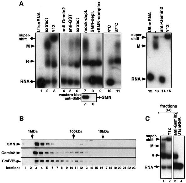
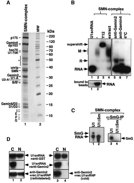
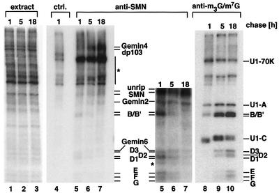
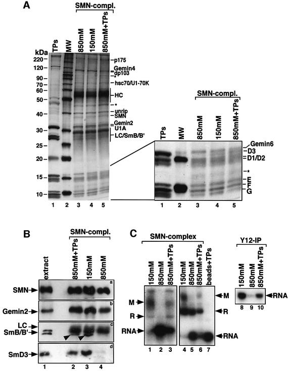
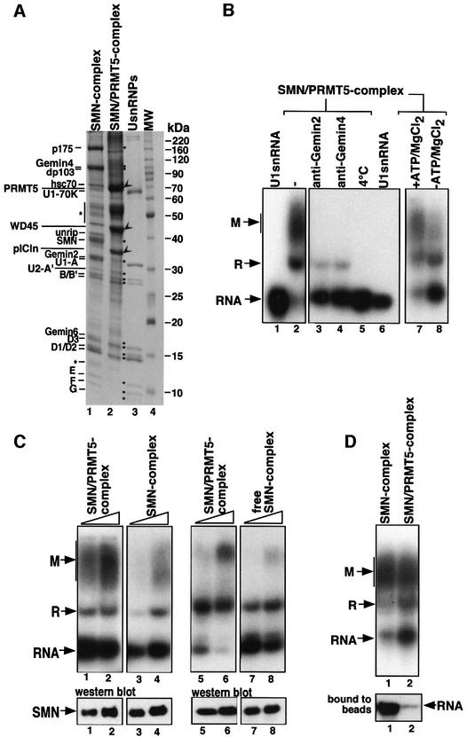
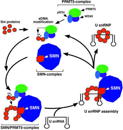
Although spliceosomal Sm proteins can assemble spontaneously onto UsnRNA in vitro, this process requires assisting factors in vivo. SMN, the protein involved in spinal muscular atrophy, is part of a complex that contains the Sm proteins and serves as a critical factor for this reaction. Here, we have reconstituted the SMN-dependent assembly of UsnRNPs in vitro. We demonstrate that the SMN complex is necessary and sufficient for the assembly reaction. The PRMT5 complex, previously implicated in methylation and storage of Sm proteins, interacts with the SMN complex and enhances its activity in an ATP-dependent manner. These data uncover the SMN-PRMT5 complex as a functional entity that promotes the assisted assembly of spliceosomal UsnRNPs, and potentially other, RNA-protein complexes.


Figures

None
Fig. 1. In vitro reconstitution of UsnRNPs in HeLa cytosolic extract. (A32P-labeled U1 snRNA (lanes 2–11) or U5 snRNA (lanes 12–15) were incubated with HeLa cytosolic extract either at 4 (lane 10) or 37°C (all other lanes). The indicated antibodies were added either after (lanes 3 and 15) or prior to assembly (lanes 4, 5 and 14). Reactions shown in lanes 7–9 were carried out in extract that was either mock depleted, SMN immunodepleted or SMN immunodepleted and subsequently incubated with purified SMN complex. The western blot below indicates the level of SMN in both extracts. Lanes 1 and 12 show snRNAs U1 and U5 in the absence of extract. The assembly reactions were separated by native gel electrophoresis and complexes visualized by autoradiography. Arrows indicate the positions of complexes R and M as well as of the Y12 supershift. (B) Fractionation of HeLa cytosolic extract active in UsnRNP assembly on a Superose-6 gel filtration column. SMN, Gemin2 and B/B′ were detected in the individual fractions by western blotting. The molecular masses of marker proteins are indicated on the top of the western blots, and fraction numbers on the bottom. (C) Fractions 3–6 were pooled and incubated with 32P-labeled U1 snRNA (lane 1) in the presence of either Y12 (added after assembly, lane 2) or Gemin2 (added prior to assembly, lane 3). Lane 4 shows the free U1 snRNA. Samples were analyzed as in (A).
None
Fig. 2. A purified SMN complex is sufficient to promote UsnRNP assembly. (A) HeLa nuclear extract was passed over an anti-SMN affinity column and proteins were analyzed by SDS–PAGE. Lane 1 shows the affinity-purified SMN complex, and lane 2 a protein size marker. Proteins of the SMN complex indicated on the left were identified by MALDI-TOF and/or western blotting. Bands indicated with an asterisk are either non-specific or have not yet been identified. (B32P-labeled U1 snRNA was incubated with Sepharose beads (lane 1) or anti-SMN affinity beads that contain the SMN complex shown in (A) (lanes 2–7). Assembly reactions in the presence of either anti-Gemin2 antibodies, anti-Gemin4 antibodies or at 4°C are shown in lanes 5, 6 and 7, respectively. The beads were pelleted and the supernatants analyzed by native gel electrophoresis as above. Y12 was added to the reaction after assembly had been completed (lane 3). The lower gel shows U1 snRNA binding to the Sepharose beads (lane 1) and to Sepharose beads containing the SMN complex (lane 2). (C) UV cross-linking of UsnRNA and the G-protein. 32P-labeled U1 snRNA (lanes 1 and 3) and U1ΔSm (lanes 2 and 4) were incubated with SMN complex for 1 h. The samples were then irradiated with UV light and either directly separated by SDS–PAGE (lanes 1 and 2) or immunoprecipitated with an anti-G antiserum (lanes 3 and 4). Bands were visualized by autoradiograpy. (D) Nuclear transport of U1 snRNP assembled by the SMN complex. 32P-labeled U1 snRNA (upper and middle panels, lanes 1–4) and U1 snRNP assembled by the SMN complex (lower panel, lanes 1 and 2) were injected into the cytoplasm of X.laevis oocytes. Oocytes were pre-injected with an anti-GST control antibody (upper panels, lanes 1–4) or with anti-Gemin2 antibody (middle and lower panels). As a further control, 32P-labeled U1 snRNA was co-injected together with cold U1 snRNP assembled by the SMN complex and Gemin2 antibodies (lower panel, lanes 3 and 4). After incubation for 12 h, RNA was extracted from the nuclear (N) or cytosolic (C) fractions, separated on a denaturing gel and visualized by autoradiography.
None
Fig. 3. Transfer of Sm proteins from the SMN complex onto UsnRNPs in vivo. HeLa cells were pulsed with [35S]methionine/[35S]cysteine for 1.5 h. After a chase with non-labeled amino acids, extracts were prepared at the indicated times and immunoprecipitated with anti-SMN antibody 7B10 (lanes 5–7), anti-m3G/m7G cap antibody H-20 (lanes 8–10) and a non-related control antibody (lane 4). Proteins were separated by SDS–PAGE and visualized by fluorography. The lower part of the anti-SMN immunoprecipitation is shown as a longer exposure. One-fiftieth of the amount of proteins used for the immunoprecipitations is shown in lanes 1–3. Bands indicated by an asterisk were immunoprecipitated non-specifically.
None
Fig. 4. An SMN complex that lacks B/B′ and D3 fails to assemble the Sm core domain. (A) Cytosolic extract active in UsnRNP assembly was incubated with an anti-SMN affinity matrix and washed with either 850 or 150 mM NaCl. Proteins bound to the matrix were eluted from the column, separated by SDS–PAGE and visualized by Coomassie Blue staining (lanes 3 and 4, respectively). In lane 5, the SMN complex was first treated with 850 mM NaCl and subsequently incubated at 150 mM NaCl with Sm proteins isolated from UsnRNPs (TPs). Unbound proteins were removed by repeated washes at 150 mM NaCl. TPs used for this procedure and a protein size marker are seen in lanes 1 and 2, respectively. A magnification of the lower part of the gel is shown on the right. The positions of co-eluting 7B10 heavy chain (HC) and light chain (LC) are indicated. (B) Western blot analysis of SMN complexes shown in (A). Monoclonal antibody 7B10 was used to detect SMN (a), and affinity-purified rabbit antisera were used to detect Gemin2 (b), B/B′ (c, indicated by arrowheads) and D3 (d). The major band seen in (c) above B/B′ corresponds to the light chain that was co-eluted from the column. (CIn vitro assembly of U1 snRNP with SMN complex isolated at 150 mM NaCl (lanes 1, 4 and 8), at 850 mM NaCl (lanes 2, 5 and 9) and after re-loading with Sm proteins (lanes 3, 6 and 10). Lane 7 shows a control assembly reaction on beads that had been incubated with TPs and was washed subsequently with PBS. Assembly of the Sm core domain was analyzed by native gel electrophoresis as above (lanes 1–7) and by immunoprecipitation using Y12 monoclonal antibody (lanes 8–10).
None
Fig. 5. Identification and characterization of an SMN–PRMT5 complex. (A) Affinity purification of the SMN complex (lane 1) and the SMN–PRMT5 complex (lane 2) on anti-SMN and anti-pICln affinity columns, respectively. Proteins that were identified by MALDI-TOF are indicated on the left. SMN complex components and the Sm proteins are marked by dots, and the PRMT5 complex proteins pICln, WD45 and PRMT5 by arrowheads. A UsnRNP protein marker and a molecular size marker were loaded in lanes 3 and 4. (B) The SMN–PRMT5 complex facilitates the ATP-dependent assembly of U1 snRNP. 32P-labeled U1 snRNA was incubated with affinity-purified SMN–PRMT5 complex (bound to the anti-pICln affinity beads, lanes 2–5 and 7–8). Anti-Gemin2 and anti-Gemin4 antibodies were added prior to the assembly reaction shown in lanes 3 and 4. In lane 8, assembly was carried out in the absence of ATP, whereas all other reactions contained 5 mM ATP. In lane 5, the SMN–PRMT5 complex was incubated with U1 snRNA at 4°C instead of 37°C; in lane 6, U1 snRNA was incubated with control beads. The beads were removed from all reactions and the supernatants analyzed by native gel electrophoresis as described in Figure 1A. (C) Increasing amounts of affinity beads containing either the SMN or the SMN–PRMT5 complex were incubated with 32P-labeled U1 snRNA. After 1 h at 37°C, the beads were pelleted and the supernatant analyzed by native gel electrophoresis (lanes 1–4, upper panel). The amount of SMN present in the assembly reactions was determined by western blotting and is shown in the lower panel. Comparison of the assembly efficiency between the SMN–PRMT5 complex (lanes 5 and 6) and the SMN complex that was dissociated from the SMN–PRMT5 complex (lanes 7 and 8). The lower western blot shows the amount of SMN in the assembly reactions. (D) Enhanced dissociation of U1 snRNA from the SMN–PRMT5 complex. In vitro assembly of U1 snRNP with either SMN complex (lane 1) or SMN–PRMT5 complex (lane 2). The RNA that remained bound to the beads in both assembly reactions is shown in the lower panel.
None
Fig. 6. A model for the SMN–PRMT5 complex-mediated assembly pathway of spliceosomal UsnRNPs. For details see text.

Similar articles

Cited by

References

    1. Brahms H., Meheus,L., de Brabandere,V., Fischer,U. and Lührmann,R. (2001) Symmetrical dimethylation of arginine residues in spliceosomal Sm protein B/B′ and the Sm-like protein LSm4 and their interaction with the SMN protein. RNA, 7, 1–12. - PMC - PubMed
    1. Bringmann P. and Luhrmann,R. (1986) Purification of the individual snRNPs U1, U2, U5 and U4/U6 from HeLa cells and characterization of their protein constituents. EMBO J., 5, 3509–3516. - PMC - PubMed
    1. Buhler D., Raker,V., Luhrmann,R. and Fischer,U. (1999) Essential role for the tudor domain of SMN in spliceosomal U snRNP assembly: implications for spinal muscular atrophy. Hum. Mol. Genet., 8, 2351–2357. - PubMed
    1. Campbell L., Hunter,K.M., Mohaghegh,P., Tinsley,J.M., Brasch,M.A. and Davies,K.E. (2000) Direct interaction of Smn with dp103, a putative RNA helicase: a role for Smn in transcription regulation? Hum. Mol. Genet., 9, 1093–1100. - PubMed
    1. Charroux B., Pellizzoni,L., Perkinson,R.A., Shevchenko,A., Mann,M. and Dreyfuss,G. (1999) Gemin3: a novel DEAD box protein that interacts with SMN, the spinal muscular atrophy gene product, and is a component of gems. J. Cell Biol., 147, 1181–1194. - PMC - PubMed

Publication types

Substances