. 2002 Nov 1;21(21):5864-74.
doi: 10.1093/emboj/cdf578.

Ribonuclease activity and RNA binding of recombinant human Dicer

Affiliations

Ribonuclease activity and RNA binding of recombinant human Dicer

Patrick Provost et al. EMBO J. .

Abstract

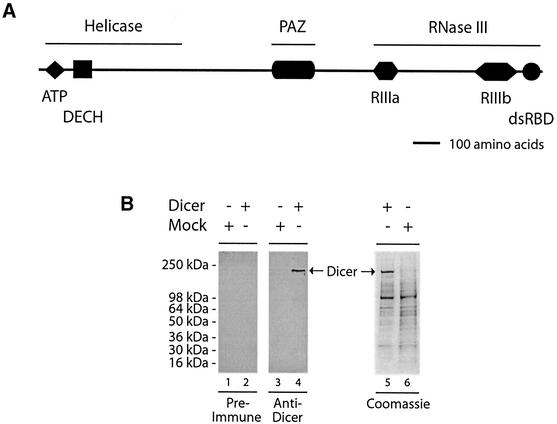
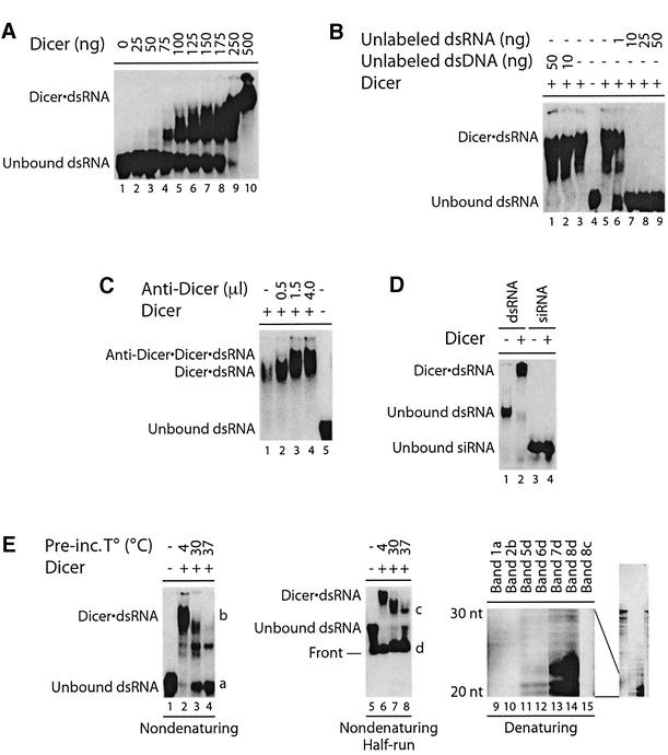
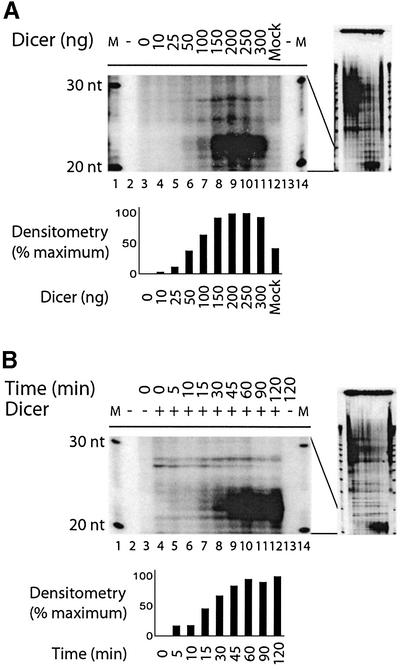
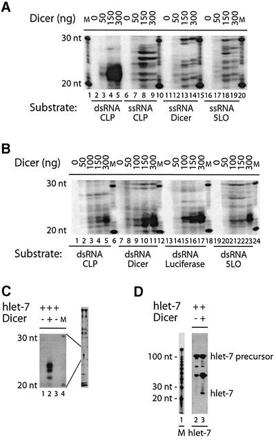
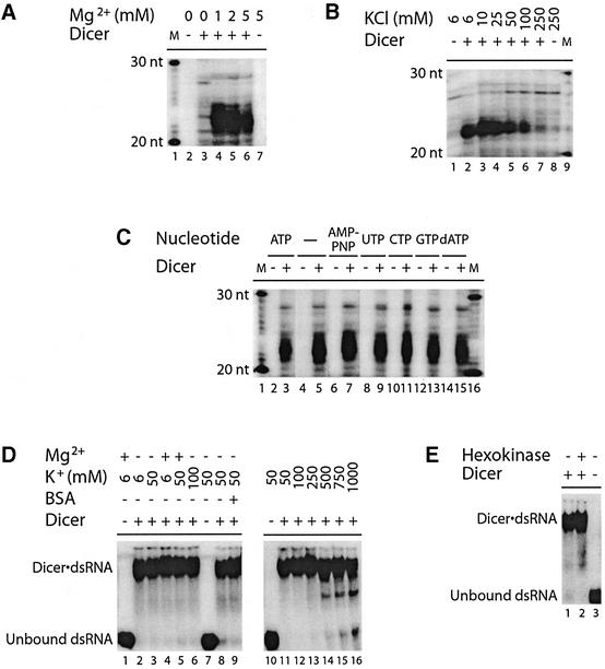
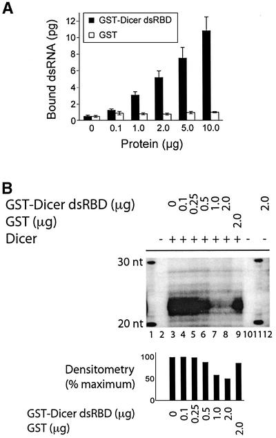
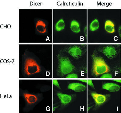
RNA silencing phenomena, known as post-transcriptional gene silencing in plants, quelling in fungi, and RNA interference (RNAi) in animals, are mediated by double-stranded RNA (dsRNA) and mechanistically intersect at the ribonuclease Dicer. Here, we report cloning and expression of the 218 kDa human Dicer, and characterization of its ribonuclease activity and dsRNA-binding properties. The recombinant enzyme generated approximately 21-23 nucleotide products from dsRNA. Processing of the microRNA let-7 precursor by Dicer produced an apparently mature let-7 RNA. Mg(2+) was required for dsRNase activity, but not for dsRNA binding, thereby uncoupling these reaction steps. ATP was dispensable for dsRNase activity in vitro. The Dicer.dsRNA complex formed at high KCl concentrations was catalytically inactive, suggesting that ionic interactions are involved in dsRNA cleavage. The putative dsRNA-binding domain located at the C-terminus of Dicer was demonstrated to bind dsRNA in vitro. Human Dicer expressed in mammalian cells colocalized with calreticulin, a resident protein of the endoplasmic reticulum. Availability of the recombinant Dicer protein will help improve our understanding of RNA silencing and other Dicer-related processes.


Figures

None
Fig. 1. Domain structure and expression of human Dicer. (A) Schematic illustration of the domain structure of the human Dicer protein. (B) Polyhistidine-tagged human Dicer protein was expressed in a baculovirus-based system, partially purified by Ni2+-affinity chromatography, and analyzed by SDS–PAGE followed by western blotting with anti-Dicer antibody or pre-immune serum, or Coomassie Blue staining. A preparation from mock-infected Sf9 cells was used as a control.
None
Fig. 2. Dicer interacts with dsRNA in EMSA. (A32P-labeled dsRNA was incubated in the absence or presence of Dicer (no MgCl2 added), (B) without or with unlabeled dsRNA or dsDNA poly(dA·dT), or (C) without or with anti-Dicer antibody (0.5–4.0 µl; no MgCl2 added). (D32P- labeled CLP 23 nt siRNA or 5LO dsRNA (100 bp) was incubated in the absence or presence of Dicer. (E) Left panel, 32P-labeled dsRNA was incubated in the absence or presence of Dicer, at the indicated temperatures for 1 h prior to EMSA analysis. Middle panel, same as left panel, but run half way. Dicer·dsRNA complex formation was analyzed by non-denaturing PAGE and autoradiography. Right panel, RNA was extracted from the indicated bands and analyzed by denaturing PAGE and autoradiography. The 20–30 nt region is shown, with the full-length gel on the right.
None
Fig. 3. Dicer is catalytically active and generates ∼21–23 nt RNA products from dsRNA in vitro. 32P-labeled CLP dsRNA (500 bp) was incubated in the absence or presence of increasing amounts of Dicer (10–300 ng), in (A) a mock preparation (300 ng) in assay buffer (pH 7.5) at 30°C for 1 h, or (B) at 30°C for the indicated time. M, indicates a 10 nt RNA size marker. The samples were analyzed by denaturing PAGE and autoradiography. The 20–30 nt regions are shown, with the full-length gels on the right. The bar graphs show quantitation of product formation by densitometric analysis of the autoradiographs, after background subtraction.
None
Fig. 4. Dicer cleaves dsRNA and miRNA let-7 precursor into ∼21–24 nt RNA products. (A32P-labeled CLP dsRNA (500 bp), or CLP ssRNA, Dicer ssRNA or 5LO ssRNA (500 nt) was incubated in the absence or presence of Dicer. (B32P-labeled CLP dsRNA, Dicer dsRNA, luciferase dsRNA or 5LO dsRNA (500 bp) was incubated in the absence or presence of Dicer. (C32P-labeled miRNA let-7 precursor was incubated in the absence or presence of Dicer. The samples were analyzed by denaturing PAGE and autoradiography. (D) miRNA let-7 precursor was incubated in the absence or presence of Dicer and analyzed by northern hybridization with a probe recognizing the mature let-7 RNA. The hybridized probe was visualized by autoradiography. M, indicates a 10 nt RNA size marker.
None
Fig. 5. Dicer dsRNase activity requires magnesium ion, but not ATP, and is sensitive to increased ionic strength. (AC) dsRNase assays. 32P-labeled CLP dsRNA (500 bp) was incubated in the absence or presence of Dicer, (A) without or with MgCl2 (1–5 mM), (B) with increasing concentrations of KCl (6–250 mM), or (C) without or with the indicated nucleotides (1 mM). M, indicates a 10 nt RNA size marker. The samples were analyzed by denaturing PAGE and autoradiography. (D and E) EMSAs. 32P-labeled 5LO dsRNA (100 bp) was incubated in the absence or presence of Dicer, (D) without or with MgCl2 (5 mM), KCl (6–1000 mM) and/or BSA (1 µg), or (E) that was pre-treated with glucose (2 mM) and/or hexokinase (0.4 U) at 30°C for 20 min prior to incubation in the absence of ATP (no KCl added). Dicer·dsRNA complex formation was analyzed by non-denaturing PAGE and autoradiography.
None
Fig. 6. The putative dsRBD of Dicer interacts with dsRNA and interferes with Dicer dsRNase activity. (A) GST-binding assay. The GST–Dicer dsRBD fusion protein, or GST alone, coupled to GSH–Sepharose 4B beads, was incubated with a 32P-labeled CLP dsRNA (500 bp) (n = 5–8 experiments). The bound 32P-labeled dsRNA was quantified by Cerenkov counting. Results are expressed as mean ± SEM. (B) dsRNase assay. 32P-labeled CLP dsRNA (500 bp) was incubated in the absence or presence of Dicer, without or with GST–Dicer dsRBD or GST. M, indicates a 10 nt RNA size marker. The samples were analyzed by denaturing PAGE and autoradiography. The bar graph shows quantitation of product formation by densitometric analysis of the autoradiographs, after background subtraction.
None
Fig. 7. Human Dicer colocalizes with the endoplasmic reticulum marker calreticulin in transiently transfected mammalian cells. (AI) Immunofluorescence microscopy. (A–C) CHO, (D–F) Cos-7, or (G–I) HeLa cells grown on coverslips were transfected with plasmid constructs encoding a Myc-tagged human Dicer. (A, D and G) Myc-Dicer was stained with anti-Myc antibody (TRITC channel, red). (B, E and H) Calreticulin was labeled with anti-calreticulin antibody (FITC channel, green). (C, F and I) Merged images demonstrates colocalization of human Dicer with calreticulin.

Similar articles

Cited by

References

    1. Abou Elela S., Igel,H. and Ares,M.,Jr (1996) RNase III cleaves eukaryotic preribosomal RNA at a U3 snoRNP-dependent site. Cell, 85, 115–124. - PubMed
    1. Bass B.L. (2000) Double-stranded RNA as a template for gene silencing. Cell, 101, 235–238. - PubMed
    1. Baulcombe D.C. (1996) RNA as a target and an initiator of post-transcriptional gene silencing in transgenic plants. Plant Mol. Biol., 32, 79–88. - PubMed
    1. Bernstein E., Caudy,A.A., Hammond,S.M. and Hannon,G.J. (2001) Role for a bidentate ribonuclease in the initiation step of RNA interference. Nature, 409, 363–366. - PubMed
    1. Billy E., Brondani,V., Zhang,H., Muller,U. and Filipowicz,W. (2001) Specific interference with gene expression induced by long, double-stranded RNA in mouse embryonal teratocarcinoma cell lines. Proc. Natl Acad. Sci. USA, 98, 14428–14433. - PMC - PubMed

Publication types