. 2002 Nov 1;21(21):5899-910.
doi: 10.1093/emboj/cdf592.

Human L1 element target-primed reverse transcription in vitro

Affiliations

Human L1 element target-primed reverse transcription in vitro

Gregory J Cost et al. EMBO J. .

Abstract

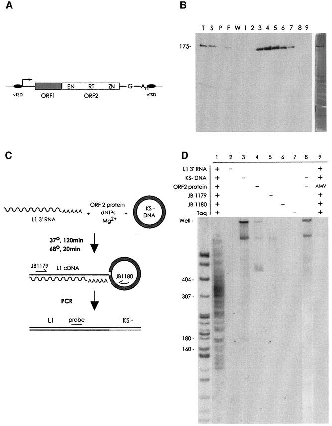
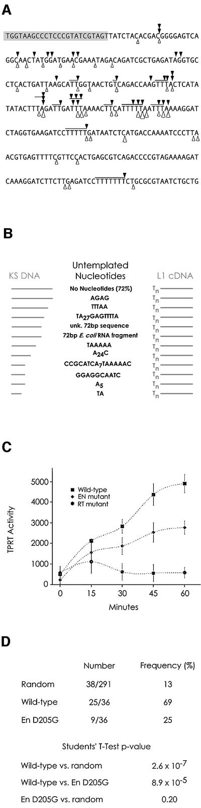
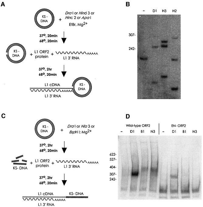
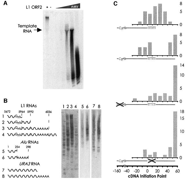
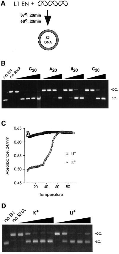
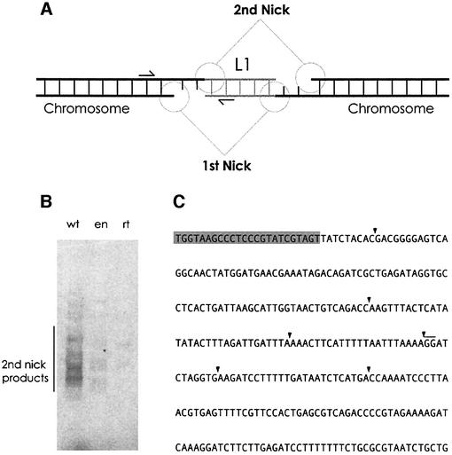
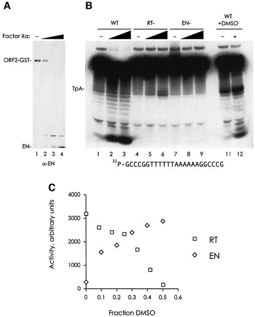
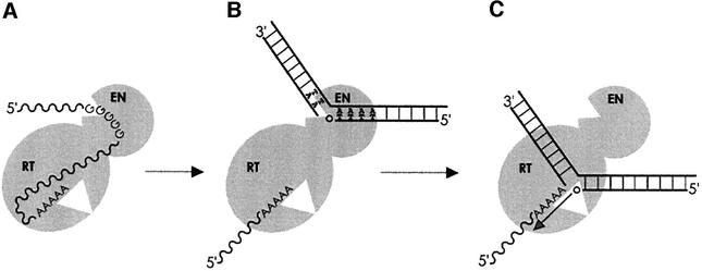
L1 elements are ubiquitous human transposons that replicate via an RNA intermediate. We have reconstituted the initial stages of L1 element transposition in vitro. The reaction requires only the L1 ORF2 protein, L1 3' RNA, a target DNA and appropriate buffer components. We detect branched molecules consisting of junctions between transposon 3' end cDNA and the target DNA, resulting from priming at a nick in the target DNA. 5' junctions of transposon cDNA and target DNA are also observed. The nicking and reverse transcription steps in the reaction can be uncoupled, as priming at pre-existing nicks and even double-strand breaks can occur. We find evidence for specific positioning of the L1 RNA with the ORF2 protein, probably mediated in part by the polyadenosine portion of L1 RNA. Polyguanosine, similar to a conserved region of the L1 3' UTR, potently inhibits L1 endonuclease (L1 EN) activity. L1 EN activity is also repressed in the context of the full-length ORF2 protein, but it and a second cryptic nuclease activity are released by ORF2p proteolysis. Additionally, heterologous RNA species such as Alu element RNA and L1 transcripts with 3' extensions are substrates for the reaction.


Figures

None
Fig. 1. (A) The human L1 retrotransposon. EN, endonuclease domain; RT, reverse transcriptase domain; ZN cysteine-rich domain; vTSD, variable target site duplication. The 5′ UTR contains an internal promoter (arrow); the 3′ UTR, a polyG and polyA sequence. (B) Protein purification. L1 ORF2p purification was analyzed by electrophoresis and western blotting and silver staining (right panel). T, total lysate; S, supernatant; P, pellet; F, column flow-through; W, wash; 1–9, 0.5 ml GSH elution fractions. (C) Reaction and detection scheme. Incubation of the reaction components results in formation of branched TPRT products. Branched molecules are detected by PCR with primers JB1179 and 1180, followed by Southern blotting with the JB2296 probe. (D) TPRT by L1 ORF2p. Lane 1, full reaction; lanes 2–7, full reaction less the indicated omission; lane 8, to ensure that the products observed in lane 1 were not the result of PCR-mediated target DNA–cDNA recombination, reactions 3 (containing cDNA but no target DNA) and 4 (containing target DNA but no cDNA) were mixed before PCR; lane 9, a full reaction, but with a large excess of AMV RT substituted for L1 ORF2p. The sizing standard used here and throughout is a MspI digest of pBR322, consisting of fragments of the following number of base pairs: 622, 527, 404, 307, 242, 238, 217, 201, 190, 180, 160, 147 and smaller fragments.
None
Fig. 2. (AIn vitro transposition insertions. L1 EN nicking sites, white arrowheads; observed L1 insertion sites, black arrowheads; JB1180 PCR primer, shaded nucleotides; ambiguity in the exact site of insertion due to microhomology between the polyT of the L1 cDNA and the target DNA, horizontal lines. (B) Untemplated nucleotides are sometimes found at transposon insertion sites. (C) Transposition activity of wild-type and mutant ORF2 proteins. Diamonds, wild-type ORF2p; squares, EN mutant ORF2p; triangles, RT mutant ORF2p. (D) Targeting of transposition. (A) contains 291 nt, 38 of which are defined as L1 target sites (see Materials and methods). Random insertion into this sequence would therefore yield an apparent targeting frequency of 13%. When wild-type L1 ORF2p was used, 25/36 L1 insertions were targeted, whereas only 9/36 insertions with EN mutant protein were. The complete set of sequenced L1 insertions exists as Supplementary information for this paper and is available from J.D.B. at http://www.bs.jhmi.edu/MBG/boekelab/boeke_lab_homepage.
None
Fig. 3. L1 TPRT can utilize pre-existing 3′ hydroxyls for transposition. (A) Pre-nicking reaction scheme. pGC89 DNA was pre-nicked with various restriction enzymes outside of the nicking and transposition hotspot region of the plasmid, then used in the TPRT reaction. (B) TPRT products are produced at the pre-nicked sites. Predicted product sizes are 252, 268 and 285, for DraI (D1 on the figure), HindIII (H3) and HincII (H2), respectively. HindIII ‘star’ nicking activity results in a band at ∼250 nt. As the pGC89 substrate used in this experiment contains four DraI sites, only 1/4th of the nicks in the plasmid are at the assayed DraI site, reducing the intensity of the DraI band four-fold relative to the other enzymes; the other sites are unique. (C) Pre-digestion of target pBluescript KS–DNA into linear fragments. (D) Transposition products are produced via utilization of either a blunt-end DSB (DraI, D1, TTT/AAA), or a four base overhang (5′ overhang, BspHI, B1, T/CATGA, all BspHI sites are also NlaIII sites; 3′ overhang, NlaIII, N3, CATG/). The region of the plasmid assayed in this experiment is within the L1 ENp nicking and L1 ORF2p TPRT hotspot region of the plasmid. Predicted molecular weights of TPRT-derived PCR products: DraI, 303 bp; NlaIII, 361 bp; BspHI, 357 bp. A band from primer–L1 cDNA fusion is seen near the bottom of all lanes in this panel. The products of the normal TPRT reaction appear lighter in this gel only because the efficiency of transposition using DSBs necessitated a shorter exposure.
None
Fig. 4. (A) In a homopolymer RT assay (polyA RNA, oligo-dT primer), L1 ORF2p produces RT products far in excess of the molecular weight of the template, indicative of template switching activity; AMV RT does not. (B) TPRT of various RNA species. The end-point of the L1 and Alu RNAs are indicated with the dotted line (not to scale). RNA number 4 has 38 nt of vector RNA after the polyA tail. URA3, fragment of S.cerevisiae URA3 RNA with and without a polyA tail. (C) Distribution of cDNA initiation points. Shown below each histogram is a full-length cDNA (from 3′ to 5′), with the positions of the reverse-transcribed polyG and polyA tracts indicated by ‘Cn’ and ‘TTTTTT’, respectively. Position zero marks the end of L1 sequence and the beginning of the polyA tail. The actual positions of the cDNA initiation are plotted in the histogram grouped into 10 nt bins. For the first cDNA (generated from RNA number 1 in B), many cDNAs end beyond the designed position at nucleotide 14 due to the addition of extra A residues to the RNA by T7 polymerase (see Materials and methods). Transposition of RNA number 4 (the second cDNA from the top) yields two distinct populations of cDNA initiation points. Mutation of either the polyG or the polyA sequence (histograms three and four, respectively) removes the bias towards internal initiation of cDNA synthesis, although only the polyA mutation results in a statistically significant difference. Student’s two-tailed t-test comparison of wild type versus mutant, bins –20–30 with bins 30–60; polyG p-value = 0.17; polyA p-value = 0.05. For all three cDNAs from 3′ extended transcripts, all initiation points in the 50–60 bin occurred at nucleotide 53, the end of the transcript. The following number of highly truncated (endpoint <–30) cDNAs were excluded from the statistical analysis: wt 3′ extended RNA, 3; polyG mutant, 4; polyA mutant, 3.
None
Fig. 5. L1 EN is inhibited by RNA. (A) L1 EN nicking activity was assayed by following conversion of the quickly migrating supercoiled KS–plasmid to the slowly migrating open-circular form. (B) L1 EN nicking of a supercoiled plasmid was challenged with 10-fold dilutions (100 µM–100 nM; 100 µM–10 nM for G20) of the indicated RNA oligo. Unlike the related CCR4 nuclease (Chen et al., 2002), L1 EN has neither polyA-specific RNA exonucleolytic activity, nor any detectable nucleolytic activity on RNA (data not shown). (C) Quartet structures (as assayed by differential light absorbance and thermal melting) are disrupted when polyI is converted to a lithium salt. (D) Quartet structures are not required for inhibition of L1 EN nicking. Ten-fold dilutions (100 ng/µl–10 pg/ml); no difference was seen even at two-fold dilutions (data not shown). sc., supercoiled; oc., open circular.
None
Fig. 6. Creation of second-strand L1 cDNA. (A) The full L1 transposition reaction requires the utilization of two nicks in the target DNA. Arrows indicate the position of the primers (JB1180 and 2NP) used for PCR. (B) Five prime end insertion. Lane 1, wild-type ORF2 protein; lane 2, EN mutant ORF2p; lane 3, RT mutant ORF2p. (C) Black arrowheads indicate the position of target DNA–L1 5′ cDNA junctions.
None
Fig. 7. L1 EN activity is masked in full-length ORF2 protein. (A) Treatment of L1 ORF2p with Factor Xa results in removal of the EN domain, as monitored by immunoblot with a L1 EN antibody. The amounts of Factor Xa used are as follows: lane 1, zero; lane 2, 0.0625 µg; lane 3, 0.25 µg; lane 4, 1 µg. (B) Proteolyzed ORF2p exhibits L1 EN activity on a double-stranded, 5′ end-labelled DNA target. The position of the TpA bond is indicated. The final concentration of DMSO added in lanes 11 and 12 was 25%. Factor Xa: lanes 1, 4, 7 and 11, zero; lanes 2, 5 and 8, 1 µg; lanes 3, 6, 9 and 12, 3 µg. (C) L1 EN activity is increased and full-length L1 ORF2p RT activity is decreased by DMSO addition. The graph for L1 EN is a quantitation and plot of the gel in Cost and Boeke (1998), figure 6a.
None
Fig. 8. The L1 TPRT model. (A) The polyA sequence positions the L1 RNA for reverse transcription; the polyG sequence could inhibit L1 EN activity. Competitive inhibition is shown for simplicity, but non-competitive inhibition could also be possible. (B) The polyG RNA may be removed from the L1 EN domain, perhaps by DNA binding or action of L1 ORF1 (Martin et al., 2000; Martin and Bushman, 2001), and L1 EN nicks the chromosome at its consensus site. (C) The nicked DNA moves to the RT active site and the newly generated 3′ hydroxyl primes reverse transcription.

Similar articles

Cited by

References

    1. Bibillo A. and Eickbush,T.H. (2002) The reverse transcriptase of the R2 non-LTR retrotransposon: continuous synthesis of cDNA on non-continuous RNA templates. J. Mol. Biol., 316, 459–473. - PubMed
    1. Boeke J.D. (1997) LINEs and Alus: the polyA connection. Nat. Genet., 16, 6–7. - PubMed
    1. Boeke J.D. and Stoye,J.P. (1997) Retrotransposons, endogenous retro viruses and the evolution of retroelements. In Coffin,J.M., Hughes,S.H. and Varmus,H.E. (eds), Retroviruses. Cold Spring Harbor Laboratory Press, Plainview, NY, pp. 343–435. - PubMed
    1. Boissinot S., Entezam,A. and Furano,A.V. (2001) Selection against deleterious line-1-containing loci in the human lineage. Mol. Biol. Evol., 18, 926–935. - PubMed
    1. Chaboissier M.C., Finnegan,D. and Bucheton,A. (2000) Retrotrans position of the I factor, a non-long terminal repeat retrotransposon of Drosophila, generates tandem repeats at the 3′ end. Nucleic Acids Res., 28, 2467–2472. - PMC - PubMed

Publication types