. 2002 Nov 1;544(3):727-39.
doi: 10.1113/jphysiol.2002.028894.

Fast synaptic transmission mediated by alpha-bungarotoxin-sensitive nicotinic acetylcholine receptors in lamina X neurones of neonatal rat spinal cord

Affiliations

Fast synaptic transmission mediated by alpha-bungarotoxin-sensitive nicotinic acetylcholine receptors in lamina X neurones of neonatal rat spinal cord

A Bradaïa et al. J Physiol. .

Abstract

Using patch clamp recordings on neonatal rat spinal cord slices, we have looked for the presence of alpha-bungarotoxin-sensitive nicotinic ACh receptors (nAChRs) on sympathetic preganglionic neurones (SPNs) surrounding the central canal of the spinal cord (lamina X) and examined whether they were implicated in a fast cholinergic synaptic transmission. SPNs were identified either by their morphology using biocytin in the recording electrode and/or by antidromic stimulation of the ventral rootlets. The selective alpha7-containing nAChR (alpha7*nAChR) agonist choline (10 mM) induced a fast, rapidly desensitizing inward current, which was fully blocked by alpha-bungarotoxin (alpha-BgT; 50 nM) and strychnine (1 microM), two antagonists of alpha7*nAChRs. The I-V relationship of the choline-induced current showed a strong inward-going rectification. Electrically evoked excitatory postsynaptic currents (eEPSCs) could be recorded. At -60 mV, eEPSCs peaked at -26.2 pA and decayed monoexponentially with a mean time constant of 8.5 ms. The current-voltage relationship for eEPSCs exhibited a strong inward rectification and a reversal potential close to 0 mV, compatible with a non-selective cationic current. The appearance of eEPSCs was entirely suppressed by the application of 100 microM ACh or nicotine. Choline (10 mM) and 1,1-dimethyl-4-phenylpiperazinium iodide (DMPP; 100 microM) both reduced the amplitude of eEPSCs, whereas cytisine (100 microM) had no effect. Strychnine (1 microM) and alpha-BgT (50 nM) both suppressed the eEPSCs. Blocking the P2X purinergic and 5-HT(3) receptors had no effect on eEPSCs. DMPP induced four types of current, which differed in their onset and desensitization rate. The most frequently encountered responses were insensitive to the action of strychnine and alpha-BgT, and were reproduced by ACh and nicotine but not by cytisine. We conclude that SPNs of the lamina X express several classes of nAChRs and in particular alpha-BgT-sensitive nAChRs. This is the first demonstration in a mammalian spinal cord preparation of a fast cholinergic neurotransmission in which alpha-BgT-sensitive nicotinic receptors are involved.


Figures
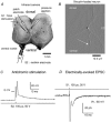
Figure 2
Figure 2. Neurone identified as a preganglionic sympathetic neurone displays fast eEPSC
A, experimental set-up. Photograph of an upper lumbar (L1-L3) transverse rat spinal cord section showing the different locations of patch electrode, electrical stimulation (S1), pressure pipette and suction stimulating electrode (S2). The recorded neurones were located laterally to the central canal (CC). The dotted white line indicates the limit of the grey matter and the central canal. B, details of the biocytin-loaded neurone revealed with FITC. Typically, neurites extend from the cell body in three directions: (a) towards the intermediate grey; (b) towards the dorsal horn; and (c) towards the ventral rootlet (indicated by the white arrow) and the ventral commissure. C, under current clamp mode, the stimulation of the ventral root whilst (bottom trace, 100 μs, -50V) using the suction electrode (S2) induced an antidromic spike in the recorded neurone. The voltage trace (top) corresponds to the average of 20 action potentials, which occurred with a constant latency. The bar indicates the 0 mV level. D, under voltage clamp at -60 mV, electrical stimulation (top trace, 100 μs, -30 V) using a focal stimulation electrode (S1) induced an EPSC (bottom trace). Recordings were made in the presence of 2 mm kynurenic acid, 10 μm CNQX and 10 μm bicuculline. ECl was fixed at -60 mV.
Figure 6
Figure 6. Effect of nicotinic receptor antagonists on DMPP-induced current
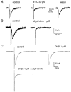
A, 100 μm DMPP was pressure-applied onto cells held at -60 mV and produced SD responses (left traces in A and B). The coapplication of 10 μm curare (d-TC) with DMPP ejected from a second pressure pipette (middle trace) induced a current of smaller amplitude. The effect of d-TC was reversible (right trace). The inhibition of the peak of the DMPP-induced current as compared to the control trace was 91 %. B, dihydro-β-erythroidine (DHβE; 10 μm) was pressure-coapplied with DMPP and reversibly (right trace) reduced (middle trace) the DMPP-induced current compared to the control trace (left trace). The inhibition of the peak of the DMPP-induced current was 34.8 % and the inhibition of the DMPP-induced current measured at the end of the application was 43 %, indicating a parallel reduction of the DMPP-induced current induced by DHβE. All traces in A and B were made at 5 min intervals. C, antagonists of α-7*nAChRs were tested in two cells showing composite responses. DMPP was pressure-applied at 100 μm and induced a biphasic current. Bath application of 50 nm α-BgT for 30 min (left traces) and bath application of 1 μm strychnine for 5 min (right traces) blocked the appearance of the fast inward current indicated by the arrows in control conditions. All recordings (A, B and C) were made in the presence of 0.5 μm TTX, 2 mm kynurenic acid and 10 μm bicuculline in neurones held at -60 mV.
Figure 7
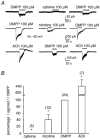
Figure 7. Relative potency of classical nicotinic receptor agonists
A, the potency of 100 μm cytisine (upper traces), 100 μm nicotine (middle traces) and 100 μm ACh (lower traces) was compared to that of 100 μm DMPP. All applications (20 s) were separated by 5 min intervals to allow recovery from desensitization. ACh was applied in the presence of 1 μm atropine, a muscarinic antagonist. Cells were held at -60 mV and recordings were made in the presence of 0.5 μm TTX, 2 mm kynurenic acid and 10 μm bicuculline. B, histogram representing the percentage of the peak of the agonist-induced current compared to that induced by DMPP in cells displaying SD and ND responses. The agonists were tested at 100 μm. The number of cells tested is given in parentheses. Error bars and * indicate the s.e.m. and significance, respectively. The apparent order of potency was: ACh >> DMPP > nicotine. Cytisine was inefficient.
Figure 1
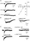
Figure 1. Choline-induced currents in neurones surrounding the central canal
A left, current traces evoked by 10 mm of choline in a neurone held at -80, -60 and 20 mV. Right, a graph showing the plot of the peak amplitude of choline-induced current (▪) versus the membrane potential. The I-V relationship shows a strong inward-going rectification as no outward current was recorded. B, current traces illustrating slow-decaying choline–induced currents. In the cell illustrated in the right panel, the slow choline-induced current was preceded by a fast inactivating current, as indicated by the arrow. C, pharmacology of the fast choline-induced current. Upper traces, 5 min bath application of 1 μm strychnine totally blocked the choline-induced current. Lower traces, pressure application of 10 mm choline evoked an inward current (left trace). Bath application of 50 nm α-bungarotoxin (α-BgT) inhibited the choline-induced current after 20 min application (right trace). All recordings (A, B and C) were made in the presence of 0.5 μm TTX, 2 mm kynurenic acid and 10 μm bicuculline. Cells in B and C were held at -60 mV.
Figure 3
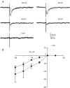
Figure 3. I-V relationship of the eEPSCs
A, eEPSCs were evoked every 3 s (100 μs, 20V) in a neurone held at holding potentials varying from -80 to 0 mV (in 20 mV steps). Each current trace represents the average of 10 eEPSCs. Recordings were made with ECl fixed at 2 mV in the presence of 10 μm bicuculline, 2 mm kynurenic acid, 10 μm CNQX and 10 μm PMBA. B, a graph showing the plot of the mean peak ± s.e.m. of eEPSCs versus potential measured in five cells with ECl at 2 mV (•). ▪, the mean peak eEPSCs ± s.e.m. measured in four cells with ECl at -60 mV. In both cases, the I-V relationships show an inward rectification as no outward eEPSCs could be recorded and a reversal potential close to 0 mV.
Figure 4
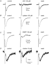
Figure 4. Cross-desensitization of eEPSCs by nicotinic receptor agonists
A, focally evoked EPSCs were recorded in neurones held at -60 mV in the presence of 2 mm kynurenic acid, 10 μm CNQX and 10 μm bicuculline. The holding potential was -60 mV and ECl was -60 mV. Stimuli (100 μs, 20 V) were delivered every 3 s. nAChR agonists were applied for 20 s using a pressure ejection pipette. Control (left panels) and wash (right panels) traces correspond to the average of 40 events before and after the application of nAChR agonists. Traces during the agonist application (middle panels) are the average of 6 events. ACh (100 μm; A) reversibly inhibited fast eEPSCs whilst cytisine (100 μm) had no effect (B). DMPP (100 μm; C) and choline 10 mm (D) reversibly reduced the current by 75 % and 42 %, respectively.
Figure 5
Figure 5. Effects of nAChR antagonists on eEPSCs
A, pressure application of 30 μmd-TC for 20 s inhibited the appearance of eEPSCs. Control and wash represent the average of 40 eEPSCs. The middle trace was obtained by averaging six traces made during the application of d-TC. Washout of the effect of d-TC was obtained 3 min after the application of d-TC. B, bath application of 1 μm strychnine entirely blocked the eEPSCs. Control and strychnine traces were obtained by averaging 40 traces before and 5 min after the beginning of application of strychnine. C, bath application of 1 μm DHβE had no effect, whilst α-BgT inhibited eEPSCs. Traces illustrate the average of 40 events before (control), during the application of 1 μm DHβE (right trace) and after 30 min application of a mixture of 50 nm α-BgT and 1 μm DHβE (bottom trace). Recordings were made at -60 mV in the presence of 2 mm kynurenic acid, 10 μm CNQX and 10 μm bicuculline. ECl was -60 mV.

References

    1. Alkondon M, Albuquerque EX. Diversity of nicotinic acetylcholine receptors in rat hippocampal neurons: I Pharmacological and functional evidence for distinct structural subtypes. Journal of Pharmacology and Experimental Therapeutics. 1993;265:1455–1473. - PubMed
    1. Alkondon M, Pereira EF, Albuquerque EX. Alpha-bungarotoxin- and methyllycaconitine-sensitive nicotinic receptors mediate fast synaptic transmission in interneurons of rat hippocampal slices. Brain Research. 1998;810:257–263. - PubMed
    1. Alkondon M, Pereira EF, Eisenberg HM, Albuquerque EX. Choline and selective antagonists identify two subtypes of nicotinic acetylcholine receptors that modulate GABA release from CA1 interneurons in rat hippocampal slices. Journal of Neuroscience. 1999;19:2693–2705. - PMC - PubMed
    1. Arias HR. Agonist self-inhibitory binding site of the nicotinic acetylcholine receptor. Journal of Neuroscience Research. 1996;44:97–105. - PubMed
    1. Barber RP, Phelps PE, Houser CR, Crawford GD, Salvaterra PM, Vaughn JE. The morphology and distribution of neurons containing acetyltransferase in the adult rat spinal cord: an immunocytochemical study. Journal of Comparative Neurology. 1984;229:329–346. - PubMed

Publication types

MeSH terms

LinkOut - more resources