. 2002 Nov 12;99(23):14758-63.
doi: 10.1073/pnas.232580699. Epub 2002 Oct 31.

The structure and evolution of the major capsid protein of a large, lipid-containing DNA virus

Affiliations

The structure and evolution of the major capsid protein of a large, lipid-containing DNA virus

Narayanasamy Nandhagopal et al. Proc Natl Acad Sci U S A. .

Abstract

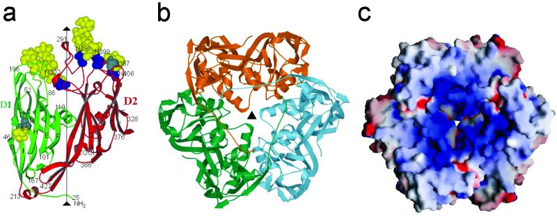
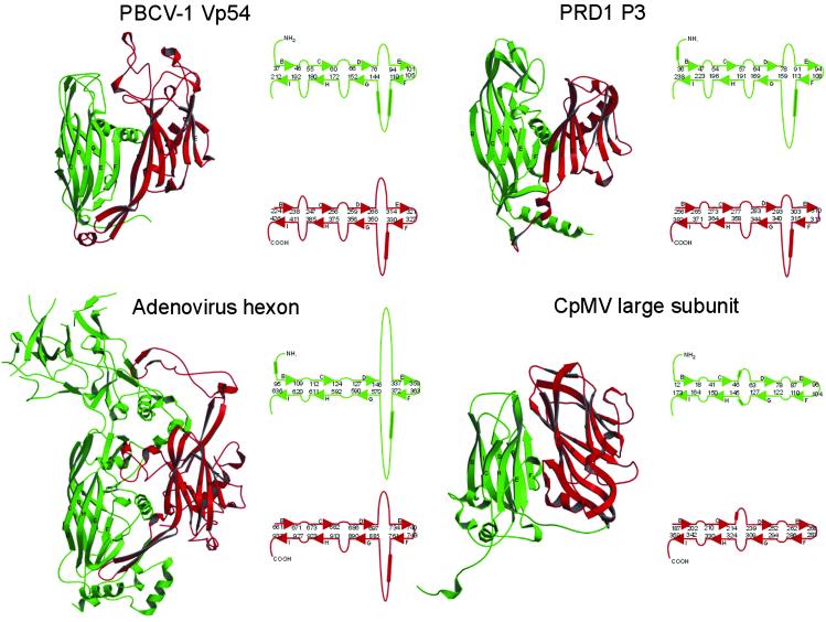
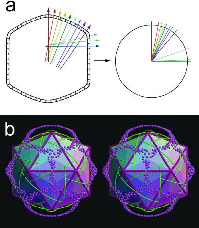
Paramecium bursaria Chlorella virus type 1 (PBCV-1) is a very large, icosahedral virus containing an internal membrane enclosed within a glycoprotein coat consisting of pseudohexagonal arrays of trimeric capsomers. Each capsomer is composed of three molecules of the major capsid protein, Vp54, the 2.0-A resolution structure of which is reported here. Four N-linked and two O-linked glycosylation sites were identified. The N-linked sites are associated with nonstandard amino acid motifs as a result of glycosylation by virus-encoded enzymes. Each monomer of the trimeric structure consists of two eight-stranded, antiparallel beta-barrel, "jelly-roll" domains related by a pseudo-sixfold rotation. The fold of the monomer and the pseudo-sixfold symmetry of the capsomer resembles that of the major coat proteins in the double-stranded DNA bacteriophage PRD1 and the double-stranded DNA human adenoviruses, as well as the viral proteins VP2-VP3 of picornaviruses. The structural similarities among these diverse groups of viruses, whose hosts include bacteria, unicellular eukaryotes, plants, and mammals, make it probable that their capsid proteins have evolved from a common ancestor that had already acquired a pseudo-sixfold organization. The trimeric capsid protein structure was used to produce a quasi-atomic model of the 1,900-A diameter PBCV-1 outer shell, based on fitting of the Vp54 crystal structure into a three-dimensional cryoelectron microscopy image reconstruction of the virus.


Figures

Fig. 1.
Fig. 1.
(a) Quasi-atomic model of the PBCV-1 capsid based on fitting the crystal structure of the Vp54 trimer into the cryo-EM reconstruction. The pentasymmetrons are colored yellow to differentiate them from the variously colored trisymmetrons. (b) Symmetry elements in the PBCV-1 trisymmetron. p3 symmetry elements are shown in black and pseudo-p6 symmetry elements are shown in gray. Pseudohexameric capsomers are presented as six disks shaded alternately in black and gray. The boundary between trisymmetrons is produced by twofold axes rotating the capsomers to face north on the left and south on the right. (c) PBCV Vp54 trimers (Cα backbone in yellow) near the center of a trisymmetron fitted into the cryo-EM map (white) viewed from outside the virus (Top) and from the side (Bottom). Figures in this paper were drawn by using the programs molscript (39), raster 3d (40), and grasp (41).
Fig. 2.
Fig. 2.
(a) Structure of the Vp54 monomer with strategic amino acids labeled. The carbohydrate moieties (yellow) and glycosylated Asn and Ser residues (blue and gray, respectively) are shown as space-filling atoms. (b) The Vp54 trimer viewed from the inside of the virus, with each monomer in a different color. (c) Surface of the Vp54 trimer also viewed from the inside of the virus, colored according to charge distribution (positive is blue, negative is red).
Fig. 3.
Fig. 3.
Comparison of virus major capsid proteins. Domains D1 and D2 are green and red, respectively. The β-sheets BIDG and CHEF are indicated. To the right of each ribbon diagram are shown diagrammatically the arrangement of β-sheets with appropriate residue numbers at the end of the β-strands. Helices are indicated by cylinders. CpMV, cowpea mosaic virus.
Fig. 4.
Fig. 4.
(a Left) A diagram representing a cut through the virion showing the surface Vp54 capsomers. The direction and position of the capsomers' threefold axes (“normals”) are shown as colored lines through each capsomer. (Right) The normals are shown shifted to a common origin. The colors of the normals in the left-hand figure correspond to the colors of the transposed normals in the right-hand figure. (b) Stereo diagram showing the normals (directions of the threefold axes) for each capsomer (purple spheres) when erected from the origin of the icosahedron as shown diagrammatically above. Boundaries between trisymmetrons and pentasymmetrons are shown in green. Icosahedral faces are shaded according to regions in which the direction of the capsomers' threefold axes lie on a common great circle.

References

    1. Yan X., Olson, N. H., Van Etten, J. L., Bergoin, M., Rossmann, M. G. & Baker, T. S. (2000) Nat. Struct. Biol. 7, 101-103. - PMC - PubMed
    1. Van Etten J. L., Lane, L. C. & Meints, R. H. (1991) Microbiol. Rev. 55, 586-620. - PMC - PubMed
    1. Tidona C. A., Schnitzler, P., Kehm, R. & Darai, G. (1998) Virus Genes 16, 59-66. - PubMed
    1. Dixon L. K., Rock, D. & Vinuela, E. (1995) in Virus Taxonomy, eds. Murphy, F. A., Fauquet, C. M., Bishop, D. H. L., Ghabrial, S. A., Jarvis, A. W., Martelli, G. P., Mayo, M. A. & Summers, M. D. (Springer, New York), pp. 92–94.
    1. Meints R. H., Lee, K., Burbank, D. E. & Van Etten, J. L. (1984) Virology 138, 341-346. - PubMed

Publication types

Substances