. 2002 Nov;161(5):1535-40.
doi: 10.1016/S0002-9440(10)64431-8.

ADAM12 alleviates the skeletal muscle pathology in mdx dystrophic mice

Affiliations

ADAM12 alleviates the skeletal muscle pathology in mdx dystrophic mice

Pauliina Kronqvist et al. Am J Pathol. 2002 Nov.

Abstract

Muscular dystrophy is characterized by muscle degeneration and insufficient regeneration and replacement of muscle fibers by connective tissue. New therapeutic strategies directed toward various forms of muscular dystrophy are needed to preserve muscle mass and promote regeneration. In this study we examined the role of the transmembrane ADAM12, a disintegrin and metalloprotease, which is normally associated with development and regeneration of skeletal muscle. We demonstrate that ADAM12 overexpression in the dystrophin-deficient mdx mice alleviated the muscle pathology in these animals, as evidenced by less muscle cell necrosis and inflammation, lower levels of serum creatine kinase, and less uptake of Evans Blue dye into muscle fibers. These studies demonstrate that ADAM12 directly or indirectly contributes to muscle cell regeneration, stability, and survival.


Figures

Figure 1.
Figure 1.
Characterization of ADAM12 transgenic mouse muscle. A–D: Immunohistochemical demonstration of endogenous and transgenic ADAM12 on frozen sections of the hindlimb muscle using a polyclonal antiserum to the C-terminal part of ADAM12 (rb 109). A: Wild-type normal mouse at embryonic day 18 (E18). B: Wild-type normal mouse at postnatal day 4. C: Wild-type normal adult mouse at 2 months. D: Adult mouse expressing transgenic ADAM12 (6 months old). E: Immunoprecipitation and Western blot of ADAM12 in transgenic (TG) mice and wild-type normal littermate controls (LC) showing the expected 110-kd proform and the 90-kd mature form of membrane-anchored ADAM12. Lanes 1 and 2 are from hindlimb muscle and lanes 3 and 4 are from diaphragm. Lane 5 represents COS-7 cells transiently transfected with a full-length human ADAM12-L expression construct as a control. A and B are cross-sections, and C and D are obliquely cut sections. Scale bars: 20 μm (A–D).
Figure 2.
Figure 2.
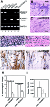
Enhanced expression of ADAM12 reduces necrosis and inflammation in the mdx mouse muscle. A: Reverse transcriptase-polymerase chain reaction analysis of mouse and human ADAM12 expression in tissue from m. quadriceps of littermate control mice (LC) (lanes 1 and 3); ADAM12 transgenic mice (TG) (lanes 2 and 4); mdx mice (lane 5); and mdx/ADAM12 mice (lane 6). Expression of GAPDH served as an internal control. Lanes 1 and 2 represent samples from E17 mice; lanes 3 to 6 are from adult (6 to 8 weeks old) mice. B: Morphological analysis of 6-week-old mdx mice quadriceps muscle with large areas of necrosis and regeneration (arrows) and of a mdx/ADAM12 muscle showing decreased pathology (C). D: Larger magnification of mdx mice and the mdx/ADAM12 quadriceps muscle (E). F and G: Immunohistochemical analysis using antibodies to the F4/80 macrophage marker (arrows) demonstrates the presence of numerous macrophages (F) in mdx muscle and a reduction of macrophages (G) in the mdx/ADAM12 mice. H: Estimate of the morphological changes (%) including necrosis, inflammation, and regenerating fibers. *, Statistically significant by the Mann-Whitney test, P < 0.0001. I: Measurements of the serum creatine kinase (U/L). Although the SD is rather big, the difference (*) between the mdx and the mdx/ADAM12 mice is statistically significant by the Mann-Whitney test at P < 0.0005. Scale bars: 60 μm (B and C); 16 μm (D and E); and 20 μm (F and G).
Figure 3.
Figure 3.
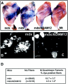
Expression of ADAM12 in the mdx mouse muscle reduces muscle necrosis. To compare the degree of muscle membrane lesions between mdx and mdx/ADAM12 mice, Evans Blue dye was injected and the hindlimbs analyzed macroscopically and by fluorescence microscopy. A:mdx mice exhibited large areas of intense blue coloration of the muscle, whereas the mdx/ADAM12 mouse exhibited much less staining, and the wild-type (Wt) mouse showed almost no coloration. B and C: Frozen sections of the quadriceps muscle were examined by fluorescence microscopy at 560 mm. D: Estimate of dye uptake. *, Statistically significant by the Mann-Whitney test at P < 0.002. Scale bars, 40 μm (B and C).

Similar articles

Cited by

References

    1. Cohn RD, Campbell KP: Molecular basis of muscular dystrophies. Muscle Nerve 2000, 23:1456-1471 - PubMed
    1. O’Brien KF, Kunkel LM: Dystrophin and muscular dystrophy: past, present, and future. Mol Genet Metab 2001, 74:75-88 - PubMed
    1. Muntoni F, Guicheney P: 85th ENMC International Workshop on Congenital Muscular Dystrophy. 6th International CMD Workshop. 1st Workshop of the Myo-Cluster Project ‘GENRE’.27–28th October 2000, Naarden, The Netherlands. Neuromuscul Disord 2002, 12:69-78 - PubMed
    1. Burton EA, Davies KE: Muscular dystrophy—reason for optimism? Cell 2002, 108:5-8 - PubMed
    1. Dubowitz V: Therapeutic possibilities in muscular dystrophy: the hope versus the hype. Neuromuscul Disord 2002, 12:113-116 - PubMed