. 2002 Nov;161(5):1577-86.
doi: 10.1016/S0002-9440(10)64436-7.

A role for caspase-1 and -3 in the pathology of experimental allergic encephalomyelitis : inflammation versus degeneration

Affiliations

A role for caspase-1 and -3 in the pathology of experimental allergic encephalomyelitis : inflammation versus degeneration

Zubair Ahmed et al. Am J Pathol. 2002 Nov.

Abstract

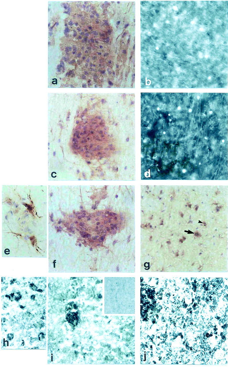
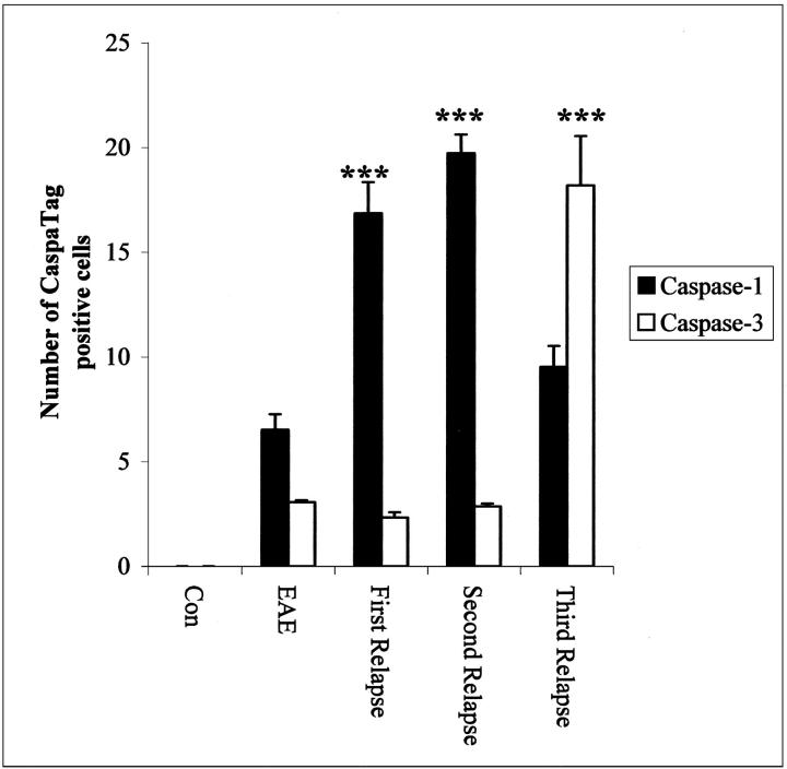
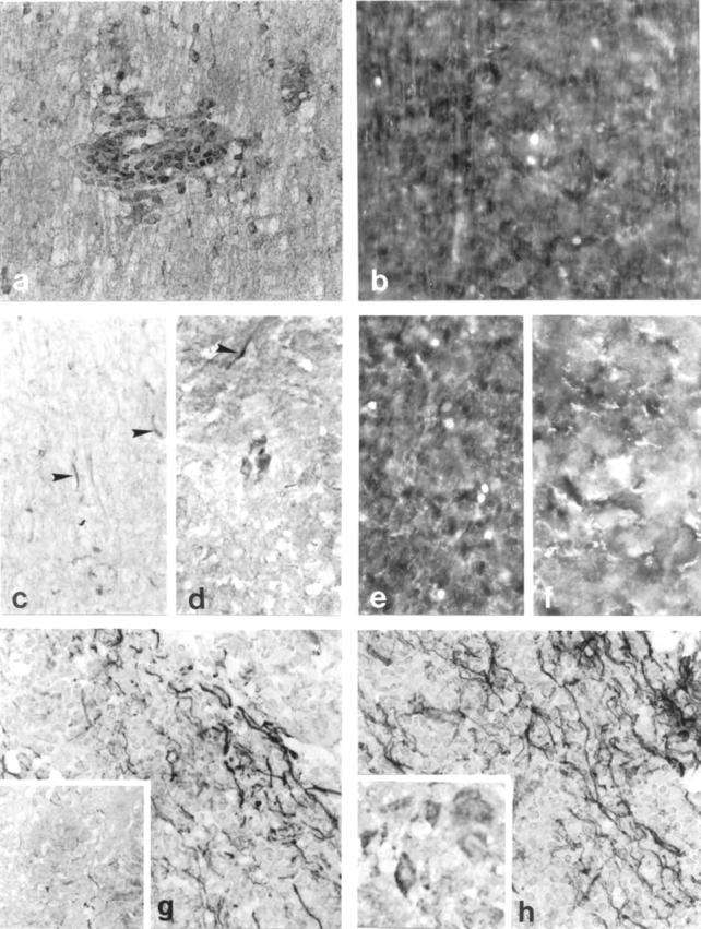
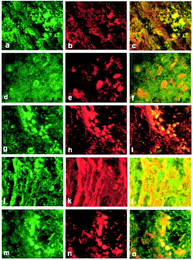
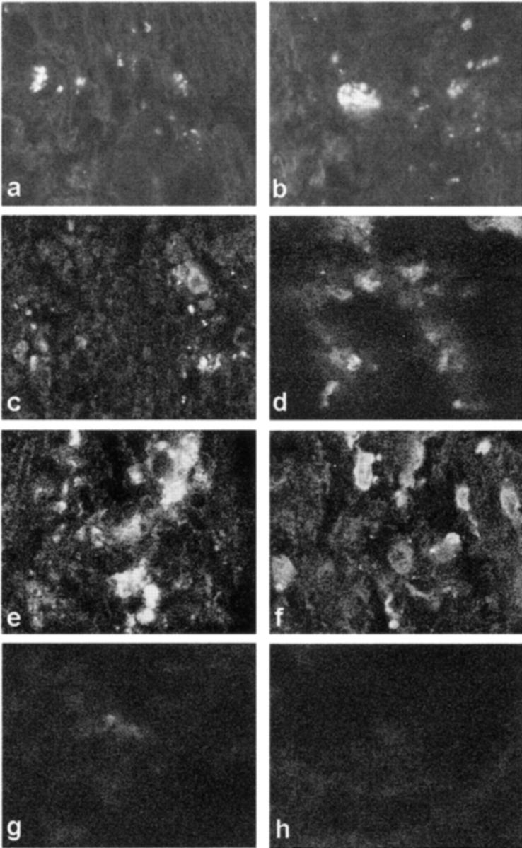
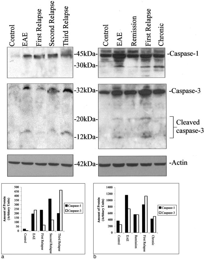
Axonal loss, already present in the acute and first relapse phases of experimental allergic encephalomyelitis (EAE) in the ABH mouse, only becomes apparent in the third relapse in the interleukin-12 model of relapsing EAE in the Lewis rat. Caspase-1 immunostaining in the spinal cord of Lewis rats was mainly localized to inflammatory cuffs with the greatest proportion of active caspase-1-positive cells detected during the first and second relapses, correlating with enzyme activity and protein on Western blots. However, in the spinal cord of ABH mice during acute EAE, caspase-1 immunostaining was localized both on inflammatory and neuronal cells, again correlating with enzyme activity and protein production. In contrast, caspase-3 expression in the spinal cord of Lewis rats did not increase significantly until the third relapse when inflammatory and neuronal cells and axons became positive in line with a significant increase in caspase activity. In ABH mice active caspase-3 was already immunolocalized on axons and apoptotic neurons in the spinal cord during the acute stage of EAE. Because caspase-3 is a downstream cell death signal it may be possible to reduce apoptosis by selectively blocking caspase-3 and therefore provide a therapeutic target for EAE and potentially, multiple sclerosis.


Figures

Figure 1.
Figure 1.
Caspase and CaspaTag-1 immunostaining. a: Spinal cord section from Lewis rat acute EAE immunostained with caspase-1. b: CaspaTag-1 immunostaining in a serial section. c: First relapse. d: Caspatag-1 in a serial section. e: Caspase-1 immunostaining in parenchymal astrocytes. f: White matter inflammatory cuff. g: Gray matter neurons of third relapse animals. h: Caspase-1-positive neurons in gray matter of ABH mice. i: Inflammatory cuff in acute EAE. j: Immunostaining in first relapse animals. Original magnifications: ×500 (a–d, f, g, i, and j); ×1000 (e and h). Inset to i shows caspase-1-negative staining in control mice.
Figure 2.
Figure 2.
Quantitation of the number of Caspatag-1 and -3+ cells in the spinal cord of Lewis rats during the relapses. ***, P < 0.0001 versus acute EAE.
Figure 3.
Figure 3.
Caspase-3 immunostaining in Lewis rat spinal cords from EAE (a), third relapse white matter (c), and third relapse (d) gray matter neuronal cells and axons (arrowhead). The corresponding CaspaTag-3 immunostaining is shown in b, e, and f, respectively. g and h: Caspase-3-immunostained axons in ABH mice acute EAE and first relapse animals, respectively. Inset to h shows caspase-3-positive immunostaining in neurons in gray matter of ABH mice. Original magnifications: ×500 (a–h); ×1000 (inset). Inset to g shows caspase-3-positive immunostaining in control mice.
Figure 4.
Figure 4.
Phenotypic characterization of active caspase-3+ cells. a and d show double staining for active caspase-3 in ED-1+ macrophages (b) and NF+ neuronal cells (e) and c and f are merged images of a and b and d and e, respectively, in the spinal cord of Lewis rats from the third relapse. g, j, and m show activated caspase-3 in CD4+ T cells (h), NF+ axons (k), and CNPase+ oligodendrocytes (n). i, l, and o are merged images of g and h, j and k, and m and n, respectively. Original magnifications, ×1500. White matter in all panels except d–f.
Figure 5.
Figure 5.
a, c, e, f, and h: White matter sections. b, d, and f: Gray matter section. a, b, and h: Lewis rat spinal cord. c–g: ABH mice. a, c, and e show fragmented DNA in inflammatory cells in white matter and b, d, and f show neuronal cells in gray matter from the spinal cords of third relapse Lewis rat and acute and relapse ABH mice, respectively. g and h: Negative control for DNA fragmentation in the spinal cord of normal animals. Original magnifications, ×1500.
Figure 6.
Figure 6.
a: Western Blot of caspase-1 and -3 protein expression during the relapses in Lewis rat EAE and a corresponding graph to show the relative amounts of protein in each band determined by gel analysis using ScionImage 4.0.2 (Scion Corporation, MD). b: Western blot in ABH mice and a graph to show the relative amounts of protein in each band. Actin blots of the same gels are shown for control of protein loading.

References

    1. Smith T, Hewson AK, Kingsley CI, Leonard JP, Cuzner ML: Interleukin-12 induces relapse in experimental allergic encephalomyelitis in the Lewis rat. Am J Pathol 1997, 150:1909-1917 - PMC - PubMed
    1. Ahmed Z, Gveric D, Pryce G, Baker D, Leonard JP, Cuzner ML: Myelin/axonal pathology in interleukin-12 induced serial relapses of experimental allergic encephalomyelitis in the Lewis rat. Am J Pathol 2001, 158:2127-2138 - PMC - PubMed
    1. Baker D, O’Neill JK, Gschmeissner SE, Wilcox CE, Butter C, Turk JL: Induction of chronic relapsing experimental allergic encephalomyelitis in Biozzi mice. J Neuroimmunol 1990, 28:261-270 - PubMed
    1. Thornberry NA, Lazebnik Y: Caspases: enemies within. Science 1998, 281:1312-1316 - PubMed
    1. Arends MJ, Wyllie AH: Apoptosis: mechanisms and roles in pathology. Int Rev Exp Pathol 1991, 32:223-254 - PubMed

Publication types

MeSH terms