. 2002 Nov;161(5):1657-67.
doi: 10.1016/S0002-9440(10)64443-4.

Expression and regulation of CD97 in colorectal carcinoma cell lines and tumor tissues

Affiliations

Expression and regulation of CD97 in colorectal carcinoma cell lines and tumor tissues

Matthias Steinert et al. Am J Pathol. 2002 Nov.

Abstract

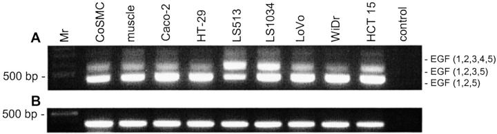
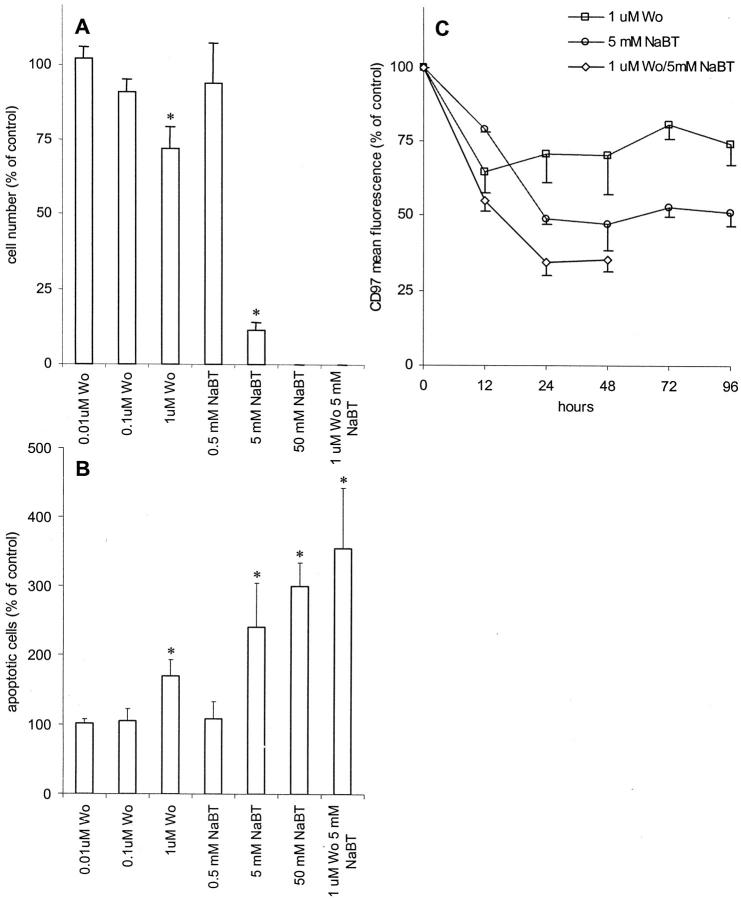
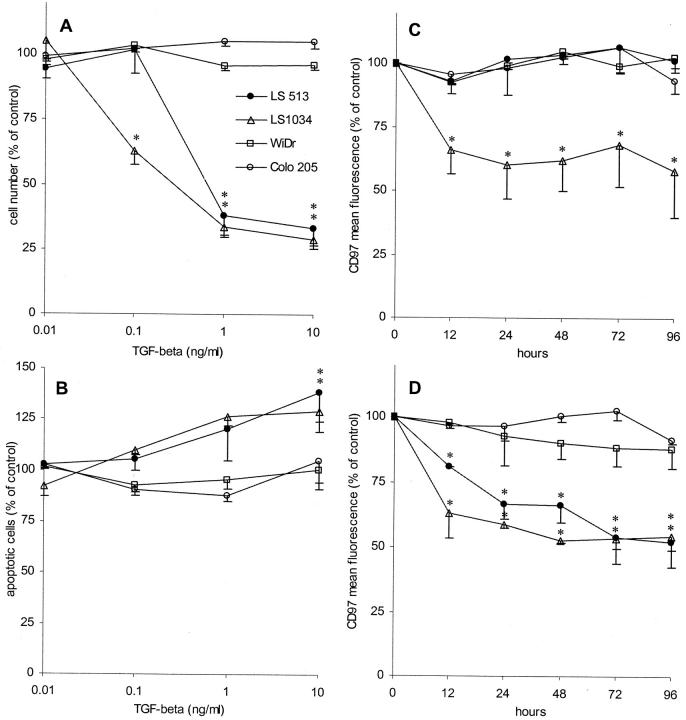
The expression of CD97, a member of the EGF-TM7 family with adhesive properties, is proportional to the aggressiveness and lymph node involvement in thyroid tumors. CD97 has never been systematically investigated in other tumors. First, we examined colorectal carcinoma cell lines (n = 18) for CD97 expression and regulation. All cell lines were CD97-positive. The level of CD97 in each line correlated with migration and invasion in vitro. This result was confirmed in CD97-inducible Tet-off HT1080 cells. Transforming growth factor-beta, which inhibits proliferation in transforming growth factor-beta-sensitive LS513 and LS1034 cells, down-regulated CD97 in these cell lines. Examining CD97 during sodium butyrate-induced cell differentiation of Caco-2 cells, we could demonstrate a CD97-decreasing effect. Second, we screened 81 colorectal adenocarcinomas by immunohistology for expression of CD97. Normal colorectal epithelium is CD97-negative. Seventy-five of 81 of the carcinomas expressed CD97. The strongest staining for CD97 occurred in scattered tumor cells at the invasion front compared to cells located within solid tumor formations of the same tumor. Carcinomas with more strongly CD97-stained scattered tumor cells showed a poorer clinical stage as well as increased lymph vessel invasion compared to cases with uniform CD97 staining. In summary, CD97 expression correlates with dedifferentiation, migration, and invasion in colorectal tumor cell lines. Moreover, more strongly CD97-stained tumor cells at the invasion front of colorectal carcinomas indicate the involvement of the molecule in tumor migration and invasion.


Figures

Figure 1.
Figure 1.
Detection of CD97 isoform mRNA by reverse transcriptase-PCR. A: The primer pair PhCD97EGFs1/r1 overspans the alternatively spliced EGF-like domain-coding region resulting in a 452-bp (EGF 1,2,5), 584-bp (EGF 1,2,3,5), or 731-bp (EGF 1,2,3,4,5) PCR product. B: Amplification of a part of the transmembrane-coding region of CD97 results in a 356-bp product. Positive control, peripheral blood lymphocytes; muscle, muscular coat of the colon; Mr, 100-bp DNA ladder.
Figure 2.
Figure 2.
Colorectal carcinoma cell lines (n = 18): correlation between CD97 expression level determined as mean fluorescence intensity in flow cytometry and percent migration (A) or percent invasion (B) determined using in vitro assays (see Table 1 ▶ ).
Figure 3.
Figure 3.
Effect of induction of differentiation in Caco-2 cells by blocking of PI3K with wortmannin or by treatment with NaBT. Results were normalized and expressed as means ± SE (n = 4) compared to the control (untreated cells). *, P ≤ 0.05 versus control. A: Effect on cell proliferation. The cells were treated with various concentrations of wortmannin (wo) and/or NaBT for 9 days. The anti-proliferative effect was evaluated in parallel using the CellTiter96AQueous one solution cell proliferation assay. B: Induction of apoptosis measured by flow cytometry. The cells were treated with various concentrations of wortmannin and/or NaBT for 48 hours. Treated and untreated cells (control) were stained with annexinV-FITC and propidium iodine. The figure demonstrates the percentage of annexin V-positive apoptotic cells. C: Effect of wortmannin and/or NaBT on the expression of CD97. Cells were treated with 1 μmol/L of wortmannin and/or 5 mmol/L of NaBT and the expression of CD97 was examined in flow cytometry.
Figure 4.
Figure 4.
TGF-β sensitivity of colorectal carcinoma cell lines. Results were normalized and expressed as means ± SE (n = 4) compared to the control (untreated cells). *, P ≤ 0.05 versus control. A: Effect of TGF-β on cell proliferation. The cells were treated with various concentrations of TGF-β for 9 days (for further details see Figure 3A ▶ ). B: Induction of apoptosis measured by flow cytometry. The cells were treated with various concentrations of TGF-β for 48 hours (for further details see Figure 3B ▶ ). C and D: Effect of TGF-β on the expression of CD97. Cells were treated with 0.1 (C) and 1 ng/ml of TGF-β (D) and the expression of CD97 was examined in flow cytometry.
Figure 5.
Figure 5.
Immunohistological expression pattern of CD97 on cryosections of colorectal normal tissue and carcinoma. A: Normal epithelial cells are CD97-negative (open arrow), whereas tumor cells express the antigen (arrow). Note the strong staining of smooth muscle cells in the lamina muscularis mucosae and the muscular coat (asterisk). B: Stronger cytoplasmic and membranous expression of CD97 in scattered tumor cells or tumor cells groups (arrow) surrounded by stroma compared to tumor cells located in tumor glands or solid tumor trabecula (open arrow). C: Stronger membranous CD97 staining of the outer parts of tumor cells at the tumor margin or invasion front (arrow) and weaker or no membrane expression on tumor cells within the center of the carcinoma. Moreover, there is a group of tumor cells seeming to detach from the tumor formation with stronger CD97 expression (open arrow). Scale bars: 100 μm (A); 50 μm (B and C).

References

    1. McKnight AJ, Gordon S: The EGF-TM7 family: unusual structures at the leukocyte surface. J Leukoc Biol 1998, 63:271-280 - PubMed
    1. Hamann J, Eichler W, Hamann D, Kerstens HM, Poddighe PJ, Hoovers JM, Hartmann E, Strauss M, van Lier RA: Expression cloning and chromosomal mapping of the leukocyte activation antigen CD97, a new seven-span transmembrane molecule of the secretion receptor superfamily with an unusual extracellular domain. J Immunol 1995, 155:1942-1950 - PubMed
    1. Gray JX, Haino M, Roth MJ, Maguire JE, Jensen PN, Yarme A, Stetler-Stevenson MA, Siebenlist U, Kelly K: CD97 is a processed, seven-transmembrane, heterodimeric receptor associated with inflammation. J Immunol 1996, 157:5438-5447 - PubMed
    1. Baud V, Chissoe SL, Viegas-Pequignot E, Diriong S, N’Guyen VC, Roe BA, Lipinski M: EMR1, an unusual member in the family of hormone receptors with seven transmembrane segments. Genomics 1995, 26:334-344 - PubMed
    1. Lin HH, Stacey M, Hamann J, Gordon S, McKnight AJ: Human EMR2, a novel EGF-TM7 molecule on chromosome 19p13.1, is closely related to CD97. Genomics 2000, 67:188-200 - PubMed

Publication types

MeSH terms