. 2002 Nov;161(5):1679-93.
doi: 10.1016/S0002-9440(10)64445-8.

Immunosuppressive treatment protects against angiotensin II-induced renal damage

Affiliations

Immunosuppressive treatment protects against angiotensin II-induced renal damage

Dominik N Muller et al. Am J Pathol. 2002 Nov.

Abstract

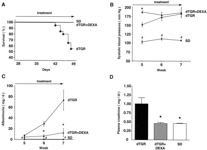
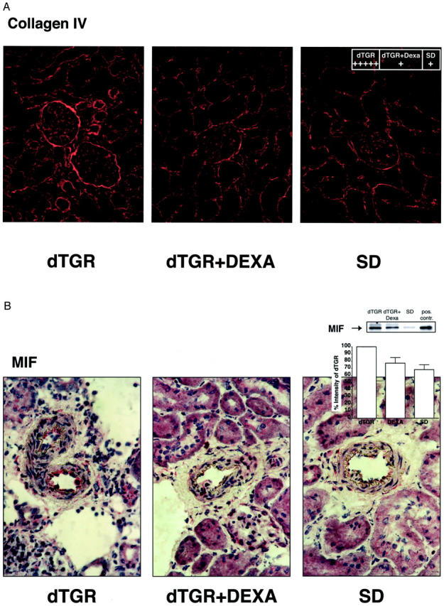
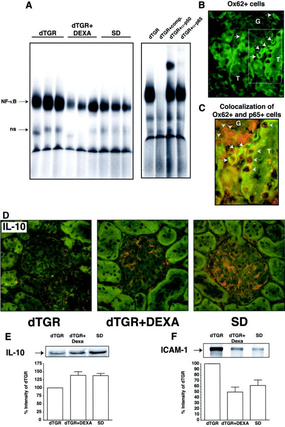
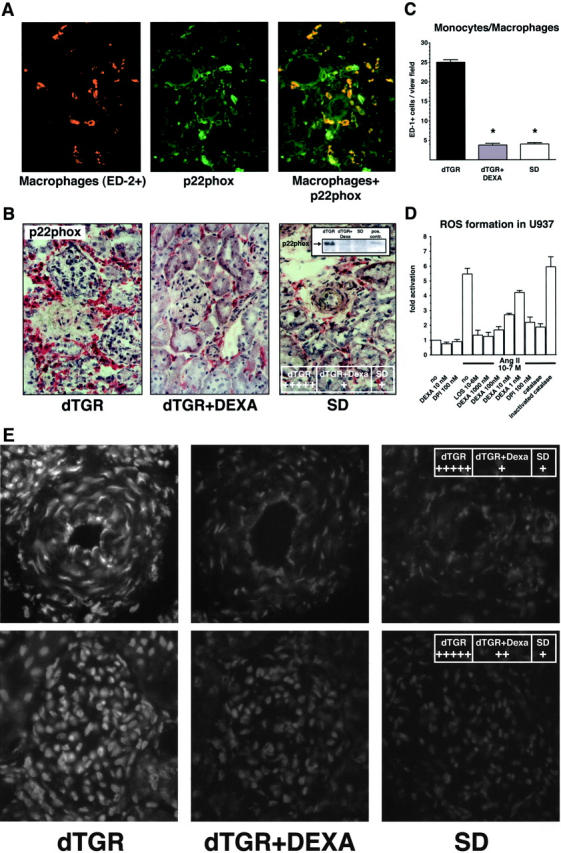
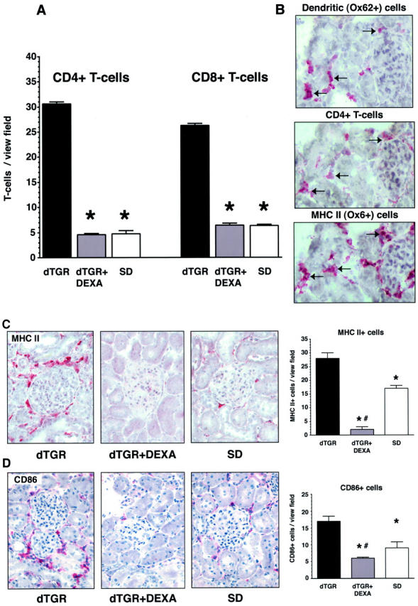
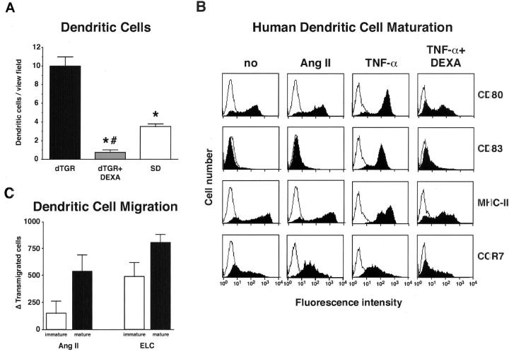
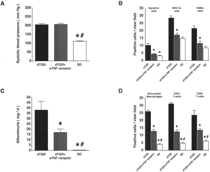
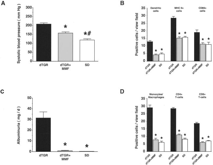
Angiotensin (Ang) II promotes renal infiltration by immunocompetent cells in double-transgenic rats (dTGRs) harboring both human renin and angiotensinogen genes. To elucidate disease mechanisms, we investigated whether or not dexamethasone (DEXA) immunosuppression ameliorates renal damage. Untreated dTGRs developed hypertension, renal damage, and 50% mortality at 7 weeks. DEXA reduced albuminuria, renal fibrosis, vascular reactive oxygen stress, and prevented mortality, independent of blood pressure. In dTGR kidneys, p22phox immunostaining co-localized with macrophages and partially with T cells. dTGR dendritic cells expressed major histocompatibility complex II and CD86, indicating maturation. DEXA suppressed major histocompatibility complex II+, CD86+, dendritic, and T-cell infiltration. In additional experiments, we treated dTGRs with mycophenolate mofetil to inhibit T- and B-cell proliferation. Reno-protective actions of mycophenolate mofetil and its effect on dendritic and T cells were similar to those obtained with DEXA. We next investigated whether or not Ang II directly promotes dendritic cell maturation in vitro. Ang II did not alter CD80, CD83, and MHC II expression, but increased CCR7 expression and cell migration. To explore the role of tumor necrosis factor (TNF)-alpha on dendritic cell maturation in vivo, we treated dTGRs with the soluble TNF-alpha receptor etanercept. This treatment had no effect on blood pressure, but decreased albuminuria, nuclear factor-kappaB activation, and infiltration of all immunocompetent cells. These data suggest that immunosuppression prevents dendritic cell maturation and T-cell infiltration in a nonimmune model of Ang II-induced renal damage. Ang II induces dendritic migration directly, whereas in vivo TNF-alpha is involved in dendritic cell infiltration and maturation. Thus, Ang II may initiate events leading to innate and acquired immune response.


Figures

Figure 1.
Figure 1.
DEXA reduced mortality and ameliorated end-organ damage independent of blood pressure in dTGRs. A: Vehicle-treated dTGRs showed >50% mortality at week 7. In contrast, no DEXA-treated dTGRs and nontransgenic SD rat died before sacrifice. B: Initially after 1 week of DEXA treatment, blood pressure increased significantly compared to vehicle-treated dTGRs. Thereafter, DEXA-treated and vehicle-treated dTGRs had similar blood pressure levels that were increased compared to SD rats. C and D: DTGR showed increased 24-hour albuminuria and plasma creatinine concentrations compared to SD rats, respectively. DEXA reduced albuminuria by 85% and normalized creatinine. Results are mean ± SEM (*, P < 0.05, dTGRs versus DEXA; #, P < 0.05, SD rats versus dTGRs and dTGRs + DEXA).
Figure 2.
Figure 2.
DEXA reduced renal fibrosis and MIF expression. A: Collagen IV in the glomerulus and glomerular basal membrane as well as peritubular area was markedly increased in dTGRs. B: MIF was markedly increased in the endothelium of damaged renal vessels as well as in infiltrated leukocytes. DEXA treatment reduced collagen IV, and MIF to control levels. Western blot of MIF from total kidney extracts confirmed the results. Rat pituitary gland extracts were used as a positive control (pos. contr.).
Figure 3.
Figure 3.
Renal NF-κB activity and co-localization of dendritic cells and p65. A: DNA-binding activity for NF-κB was markedly induced in dTGRs. Nontransgenic SD rats showed no NF-κB activity. DEXA reduced NF-κB activity toward SD rats. Competition and supershift experiments with antibodies against p50 and p65 showed specificity of the NF-κB activity. B: Renal Ox62+ dendritic cell infiltration (green staining indicated by white arrows). C: A higher magnification demonstrated that dendritic cells (green) co-localized with nuclear p65 staining (red) marked by white arrows. Tubules (T) and glomeruli (G) of vehicle-treated dTGRs showed also nuclear p65 staining (gray arrowheads). D: IL-10 was expressed in glomeruli of DEXA-treated and nontransgenic rats. E and F: Representative Western blot analysis of total kidney extracts confirmed the IL-10 and ICAM-1 immunohistochemical results, respectively.
Figure 4.
Figure 4.
Macrophage infiltration, p22phox NAD(P)H subunit, and in situ ROS production in renal sections. A: Co-localization of macrophages (red) and p22phox (green) and the merged picture (orange). Almost all perivascular-infiltrated macrophages express p22phox. B: Representative photomicrograph of p22phox showed increased expression in dTGRs because of infiltrated leukocytes. DEXA reduced expression levels to SD rat controls. Representative p22phox Western blot from total kidney extracts confirmed the results. TNF-α-treated leukocytes were used as positive control (pos. contr.). C: Semiquantification of renal sections revealed that DEXA reduced infiltrated monocytes/macrophages (ED-1+) to nontransgenic level. D: Fluorescence signal of U937 macrophages preincubated with DCFH-DA was directly induced by Ang II. Preincubation with catalase (1000 U/ml), diphenylene iodonium (10 μmol/L), and DEXA (1 to 1000 nmol/L) inhibited the Ang II-induced fluorescence intensity. None of the inhibitors changed the baseline of ROS formation. E: Vehicle-treated dTGRs show a fluorescent ethidium signal in the endothelium, smooth muscle cells, adventitia of the vessel wall, infiltrated cells, as well as in the glomerulus, which was markedly attenuated by DEXA.
Figure 5.
Figure 5.
DEXA suppresses immune cells. A: Vehicle-treated dTGR kidneys showed a significantly increased infiltration of CD4+ and CD8+ T cells. DEXA-treated and SD rats showed a similar number of CD4+ and CD8+ T cells. B: Serial kidney sections (6 μm) of vehicle-treated dTGRs demonstrated that dendritic cells (top) express MHC II (bottom) and are adjacent to CD4+ cells (middle) indicated by the black arrows. C and D: Vehicle-treated dTGRs show perivascular and periglomerular infiltration of MHC II+ and CD86+ cells, respectively. DEXA reduced MHC II and CD86 expression below nontransgenic level.
Figure 6.
Figure 6.
Effect of Ang II on dendritic cell in vivo and in vitro. A: Vehicle-treated dTGRs showed increased dendritic cell infiltration into the kidney that was reduced even below SD rat level by DEXA. B: FACS analysis of human dendritic cells showed that Ang II was unable to promote maturation. In contrast, TNF-α induced dendritic cell maturation. This effect was blocked by DEXA. In addition, Ang II and TNF-α both induced CCR7 expression. C: Chemotaxis assay of immature dendritic cells at day 7 of differentiation and mature dendritic cells. For maturation, cells were cultured at day 6 for 24 hours with TNF-α. Both, Ang II and Epstein-Barr virus-induced molecule 1 ligand chemokine, the ligand for CCR7 as positive control, increased cell migration. Cell migration was more pronounced in mature, compared to immature dendritic cells.
Figure 7.
Figure 7.
Chronic treatment with a soluble TNF-α receptor (sTNF receptor) reduced macrophage, dendritic, and T-cell infiltration; expression of MHC II and CD86; and ameliorated renal damage independent of blood pressure in dTGRs. A: Systolic blood pressure was not affected by sTNF receptor treatment. B: In contrast, sTNF receptor treatment reduced albuminuria. C: Infiltration of dendritic, MHC II+, and CD86+ cells in the kidney was reduced by sTNF receptor treatment toward SD rat level. D: In contrast, macrophage and T-cell infiltration was partially reduced. Results are mean ± SEM (*, P < 0.05, dTGR versus dTGR + sTNF receptor and SD; #, P < 0.05 SD versus dTGR + sTNF receptor).
Figure 8.
Figure 8.
Chronic MMF treatment reduced macrophage, dendritic, and T-cell infiltration; expression of MHC II and CD86; and ameliorated renal damage. A and B: MMF partially reduced systolic blood pressure, whereas albuminuria was normalized. C and D: Infiltration of dendritic, MHC II+, CD86+, and T cells, and macrophages in the kidney was reduced by MMF treatment toward SD rat level. Results are mean ± SEM (*, P < 0.05, dTGR versus dTGR + MMF and SD; *, P < 0.05 SD versus dTGR + MMF).

References

    1. Rodriguez-Iturbe B, Pons H, Herrera-Acosta J, Johnson RJ: Role of immunocompetent cells in nonimmune renal diseases. Kidney Int 2001, 59:1626-1640 - PubMed
    1. Lombardi D, Gordon KL, Polinsky P, Suga S, Schwartz SM, Johnson RJ: Salt-sensitive hypertension develops after short-term exposure to Angiotensin II. Hypertension 1999, 33:1013-1019 - PubMed
    1. Mai M, Geiger H, Hilgers KF, Veelken R, Mann JF, Dammrich J, Luft FC: Early interstitial changes in hypertension-induced renal injury. Hypertension 1993, 22:754-765 - PubMed
    1. Grafe M, Auch-Schwelk W, Zakrzewicz A, Regitz-Zagrosek V, Bartsch P, Graf K, Loebe M, Gaehtgens P, Fleck E: Angiotensin II-induced leukocyte adhesion on human coronary endothelial cells is mediated by E-selectin. Circ Res 1997, 81:804-811 - PubMed
    1. Krejcy K, Eichler HG, Jilma B, Kapiotis S, Wolzt M, Zanaschka G, Gasic S, Schutz W, Wagner O: Influence of angiotensin II on circulating adhesion molecules and blood leukocyte count in vivo. Can J Physiol Pharmacol 1996, 74:9-14 - PubMed

MeSH terms

Substances