. 2002 Nov;161(5):1749-59.
doi: 10.1016/S0002-9440(10)64452-5.

Guanylate-binding protein-1 expression is selectively induced by inflammatory cytokines and is an activation marker of endothelial cells during inflammatory diseases

Affiliations

Guanylate-binding protein-1 expression is selectively induced by inflammatory cytokines and is an activation marker of endothelial cells during inflammatory diseases

Clara Lubeseder-Martellato et al. Am J Pathol. 2002 Nov.

Abstract

During angiogenesis and inflammatory processes, endothelial cells acquire different activation phenotypes, whose identification may help in understanding the complex network of angiogenic and inflammatory interactions in vivo. To this goal we investigated the expression of the human guanylate-binding protein (GBP)-1 that is highly induced by inflammatory cytokines (ICs) and, therefore, may characterize IC-activated cells. Using a new rat monoclonal antibody raised against GBP-1, we show that GBP-1 is a cytoplasmic protein and that its expression in endothelial cells is selectively induced by interferon-gamma, interleukin-1alpha, interleukin-1beta, or tumor necrosis factor-alpha, but not by other cytokines, chemokines, or growth factors. Moreover, we found that GBP-1 expression is highly associated with vascular endothelial cells as confirmed by the simultaneous detection of GBP-1 and the endothelial cell-associated marker CD31 in a broad range of human tissues. Notably, GBP-1 expression was undetectable in the skin, but it was highly induced in vessels of skin diseases with a high-inflammatory component including psoriasis, adverse drug reactions, and Kaposi's sarcoma. These results indicate that GBP-1 is a novel cellular activation marker that characterizes the IC-activated phenotype of endothelial cells.


Figures

Figure 1.
Figure 1.
Selective induction of GBP-1 expression in endothelial cells by ICs. A: Purified recombinant GBP-1, GBP-2, and eGFP (100 ng each) were detected by Western blot using mAb 1B1 and mAb 6F12. B: Western blot analysis of GBP-1 expression in different cell types that were either untreated (−) or stimulated with IFN-γ (100 U/ml) for 16 hours. C: Western blot analysis of GBP-1 expression in HUVECs stimulated with the indicated factors for 24 hours. The following concentrations were used: IFN-γ, 100 U/ml; IL-1α, 5 ng/ml; IL-1β, 200 U/ml; TNF-α, 300 U/ml; IL-4, 10 U/ml; IL-6, 50 U/ml; IL-10, 50 ng/ml; IL-18, 100 ng/ml; oncostatin M, 10 ng/ml; MCP-1, 50 ng/ml; PF4, 25 ng/ml; SDF-1α, 200 ng/ml; bFGF, 10 ng/ml; VEGF, 10 ng/ml; Ang-2, 800 ng/ml; and PDGF B/B, 100 ng/ml. It was confirmed by our experiments and it is according to the literature that in the concentrations used each factor induces a significant cell biological response in these cells. In addition IP-10 (50 ng/ml) was used in a concentration similar to that established in the supernatant of IFN-γ-stimulated endothelial cells. MIP-1β (50 ng/ml) in the concentration used exerts maximal chemotactic activity on human T cells. mAb 1B1 was used in B and C. Immunochemical detection of actin (Actin) demonstrated that equal amounts of proteins were blotted onto the membranes.
Figure 2.
Figure 2.
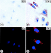
Localization of GBP-1 in the cytoplasm of endothelial cells. GBP-1 expression in HUVECs was analyzed by standard ABC immunoperoxidase staining (A, B) or by indirect immunofluorescence (C, D) using mAb 1B1 before (A, C) or after (B, D) stimulation with 100 U/ml IFN-γ for 16 hours. Original magnifications: ×450 (A, B); ×630 (C, D).
Figure 3.
Figure 3.
Expression of GBP-1 in blood vessels of various human tissues. Detection of GBP-1 expression using the standard ABC immunohistochemical method in paraffin sections of spleen (A, I), uterus (B), lung (C), heart (D), and skin (E, G). Controls: preadsorption of mAb 1B1 with an excess (300-fold) of purified GBP-1-His (G, lung) and staining with an isotypic control antibody (H, spleen); mAb 1B1 was used in A–F and mAb 6F12 was used in I. Examples of GBP-1-positive (black arrows) and -negative (white arrows) vessels are indicated. Original magnifications, ×250.
Figure 4.
Figure 4.
Expression of GBP-1 in vascular endothelial cells in human tissues. Indirect immunofluorescence staining of tissue sections of bladder (A), endometrium (B), heart (C), and lung (D) for GBP-1 (green, left) and the endothelial cell-associated antigen CD31 (red, middle). Merging of the two pictures (right) shows co-localization of GBP-1 and CD31 (yellow, white arrows). Original magnifications, ×400.
Figure 5.
Figure 5.
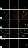
Induction of GBP-1 expression in vascular endothelial cells in diseases of the skin with a high-inflammatory component. Indirect immunofluorescence staining of tissue sections of healthy skin (A), KS (B, E), adverse drug reaction of the skin (C), and psoriasis (D) for GBP-1 (green, left, A–E) and the endothelial cell-associated antigen CD31 (red, middle, A–D). Merging of the two pictures (right) shows co-localization (yellow, white arrows). Original magnifications: ×250 (A, B, E); ×400 (C, D).

Similar articles

Cited by

References

    1. Cines DB, Pollak ES, Buck CA, Loscalzo J, Zimmerman GA, McEver RP, Pober JS, Wick TM, Konkle BA, Schwartz BS, Barnathan ES, McCrae KR, Hug BA, Schmidt AM, Stern DM: Endothelial cells in physiology and in the pathophysiology of vascular disorders. Blood 1998, 91:3527-3561 - PubMed
    1. Pober JS: Warner-Lambert/Parke-Davis award lecture. Cytokine-mediated activation of vascular endothelium. Physiology and pathology. Am J Pathol 1988, 133:426-433 - PMC - PubMed
    1. Cotran RS, Pober JS: Endothelial Activation: Its Role in Inflammatory and Immune Reactions. 1988:pp 335-347 Plenum Press, New York
    1. Roesen P, Ferber P, Tschoepe D: Macrovascular disease in diabetes: current status. Exp Clin Endocrinol Diabetes 2001, 109(Suppl):S474-S486 - PubMed
    1. Baumgartl HJ, Standl E: The atherosclerotic process and its exacerbation by diabetes. Exp Clin Endocrinol Diabetes 2001, 109(Suppl):S487-S492 - PubMed

Publication types

MeSH terms