

Aldosterone-induced inflammation in the rat heart : role of oxidative stress
- PMID: 12414524
- PMCID: PMC1850792
- DOI: 10.1016/S0002-9440(10)64454-9
Aldosterone-induced inflammation in the rat heart : role of oxidative stress
Abstract
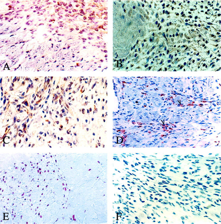
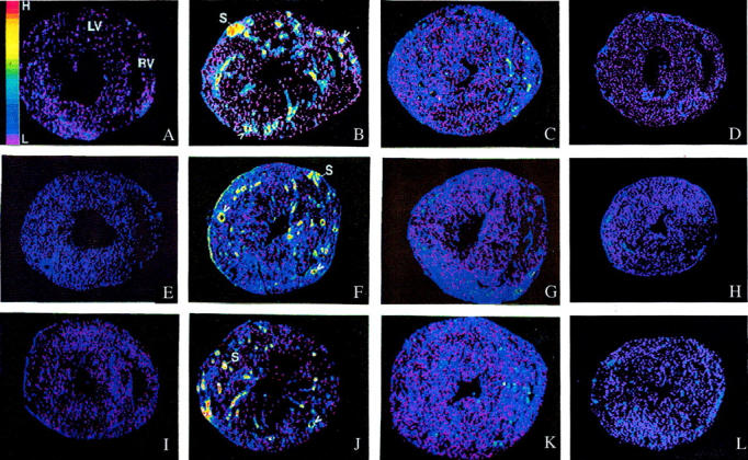
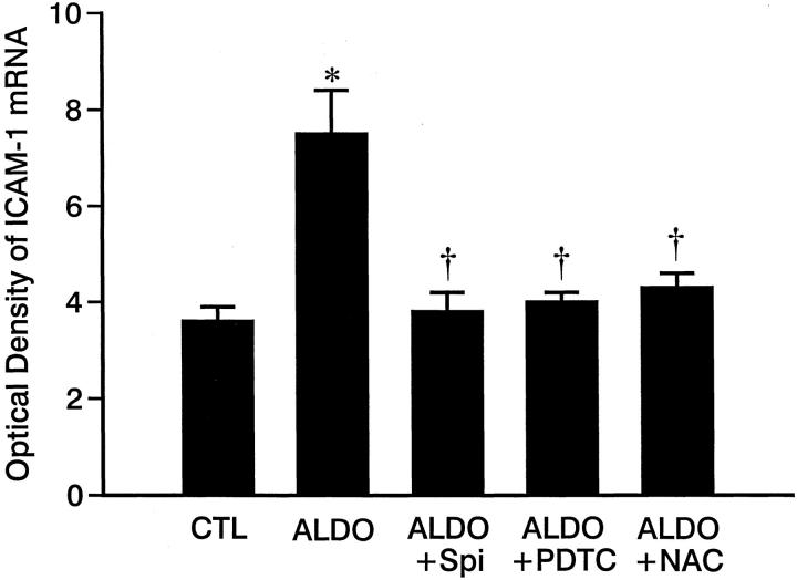
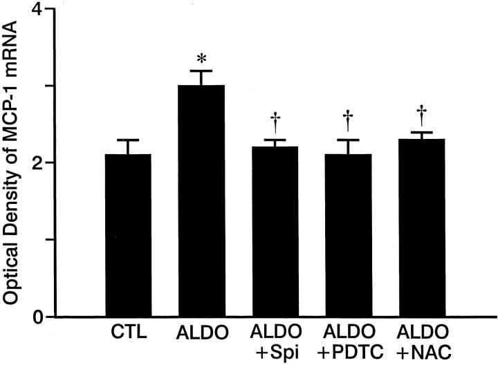
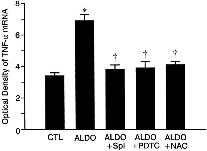
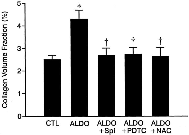
Heart failure and hypertension have each been linked to an induction of oxidative stress transduced by neurohormones, such as angiotensin II and catecholamines. Herein, we hypothesized that aldosterone (ALDO) likewise induces oxidative stress and accounts for a proinflammatory/fibrogenic phenotype that appears at vascular and nonvascular sites of injury found in both right and left ventricles in response to ALDO/salt treatment and that would be sustained with chronic treatment. Uninephrectomized rats received ALDO (0.75 micro g/hour) together with 1% dietary NaCl, for 3, 4, or 5 weeks. Other groups received this regimen in combination with an ALDO receptor antagonist, spironolactone (200 mg/kg p.o. daily), or an antioxidant, either pyrrolidine dithiocarbamate (PDTC) (200 mg/kg s.c. daily) or N-acetylcysteine (NAC) (200 mg/kg i.p. daily). Unoperated and untreated age- and gender-matched rats served as controls. We monitored spatial and temporal responses in molecular and cellular events using serial, coronal sections of right and left ventricles. Our studies included: assessment of systolic blood pressure; immunohistochemical detection of NADPH oxidase expression and activity; analysis of redox-sensitive nuclear factor-kappaB activation; in situ localization of intercellular adhesion molecule-1, monocyte chemoattractant protein-1, and tumor necrosis factor-alpha mRNA expression; monitoring cell growth and infiltration of macrophages and T cells; and analysis of the appearance and quantity of fibrous tissue accumulation. At week 3 of ALDO/salt treatment and comparable to controls, there was no evidence of oxidative stress or pathological findings in the heart. However, at weeks 4 and 5 of treatment, increased gp91(phox) and 3-nitrotyrosine expression and persistent activation of RelA were found in endothelial cells and inflammatory cells that appeared in the perivascular space of intramural coronary arteries and at sites of lost cardiomyocytes in both ventricles. Coincident in time and space with these events was increased mRNA expression of intercellular adhesion molecule-1, monocyte chemoattractant protein-1, and tumor necrosis factor-alpha. Macrophages, lymphocytes, and proliferating endothelial and vascular smooth muscle cells and fibroblast-like cells were seen at each of these sites, together with an accumulation of fibrillar collagen, or fibrosis, as evidenced by a significant increase in ventricular collagen volume fraction. Co-treatment with spironolactone, PDTC, or NAC attenuated these molecular and cellular responses as well as the appearance of fibrosis at vascular and nonvascular sites of injury. Furthermore, elevated systolic blood pressure in ALDO-treated rats was partially suppressed by spironolactone or either antioxidant. Thus, chronic ALDO/salt treatment is accompanied by a time-dependent sustained activation of NADPH oxidase with 3-nitrotyrosine generation and nuclear factor-kappaB activation expressed by endothelial cells and inflammatory cells. This leads to a proinflammatory/fibrogenic phenotype involving vascular and nonvascular sites of injury found, respectively, in both normotensive and hypertensive right and left ventricles. Spionolactone, PDTC, and NAC each attenuated these responses suggesting ALDO/salt induction of oxidative/nitrosative stress is responsible for the appearance of this proinflammatory phenotype.
Figures
References
-
- Weglicki WB, Kramer JH, Mak IT: The role of antioxidant drugs in oxidative injury of cardiovascular tissue. Heart Fail Rev 1999, 4:183-192
-
- Ball AM, Sole MJ: Oxidative stress and the pathogenesis of heart failure. Cardiol Clin 1998, 16:665-675 - PubMed
-
- Keith ME, Jeejeebhoy KN, Langer A, Kurian R, Barr A, O’Kelly B, Sole MJ: A controlled clinical trial of vitamin E supplementation in patients with congestive heart failure. Am J Clin Nutr 2001, 73:219-224 - PubMed
-
- Sole MJ, Jeejeebhoy KN: Conditioned nutritional requirements and the pathogenesis and treatment of myocardial failure. Curr Opin Clin Nutr Metab Care 2000, 3:417-424 - PubMed
-
- Singal PK, Khaper N, Farahmand F, Bello-Klein A: Oxidative stress in congestive heart failure. Curr Cardiol Rep 2000, 2:206-211 - PubMed
Publication types
MeSH terms
Substances
Grants and funding
LinkOut - more resources
Full Text Sources
Other Literature Sources
Medical
Research Materials
