Comparative Study
. 2002 Nov;161(5):1783-96.
doi: 10.1016/S0002-9440(10)64455-0.

Fas and fas ligand are up-regulated in pulmonary edema fluid and lung tissue of patients with acute lung injury and the acute respiratory distress syndrome

Affiliations
Comparative Study

Fas and fas ligand are up-regulated in pulmonary edema fluid and lung tissue of patients with acute lung injury and the acute respiratory distress syndrome

Kurt H Albertine et al. Am J Pathol. 2002 Nov.

Abstract

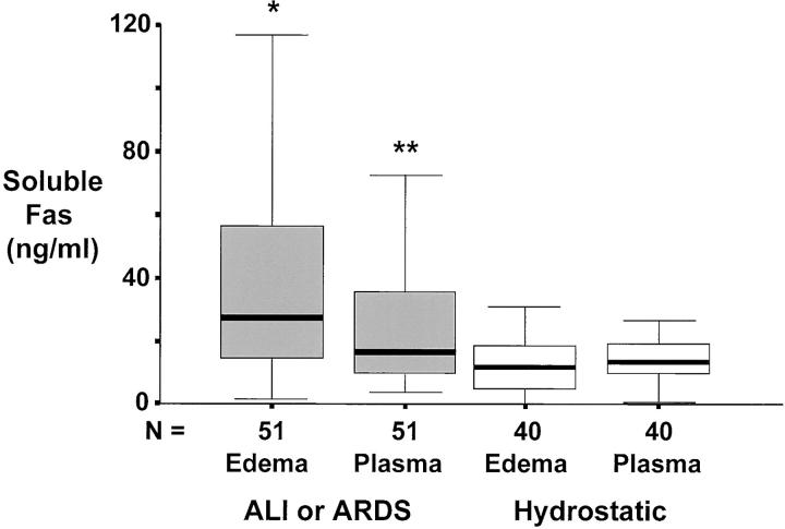
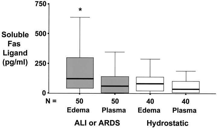
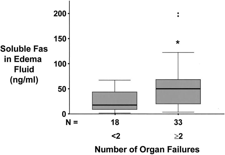
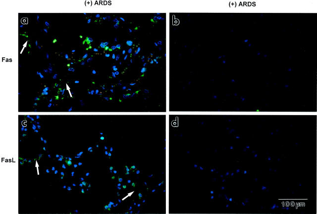
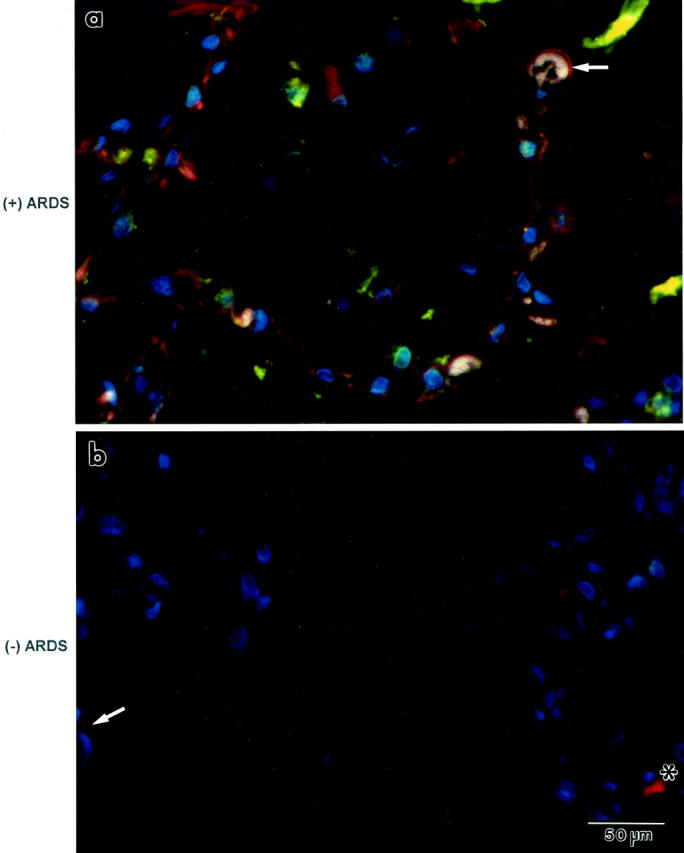
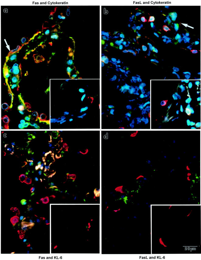
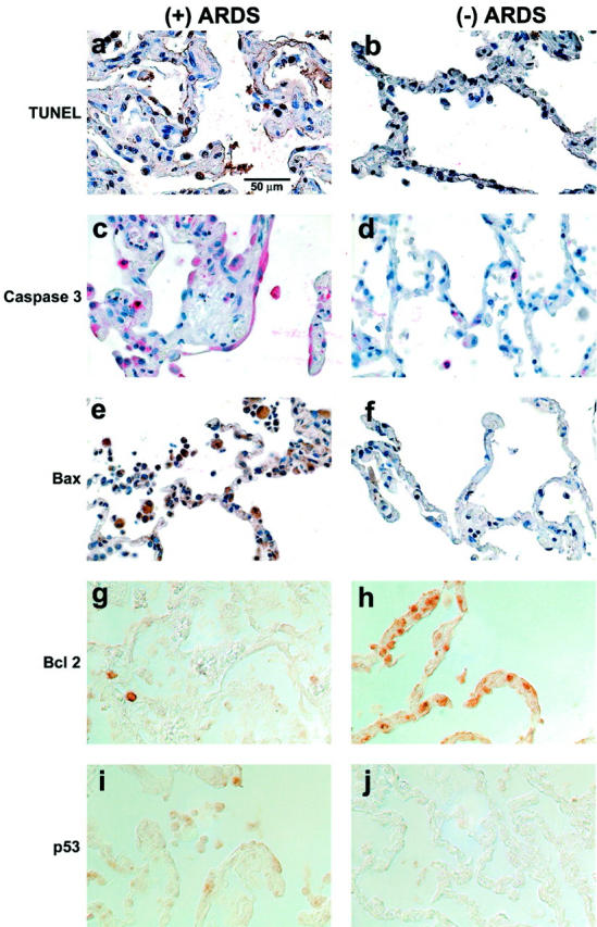
Apoptosis mediated by Fas/Fas ligand (FasL) interaction has been implicated in human disease processes, including pulmonary disorders. However, the role of the Fas/FasL system in acute lung injury (ALI) and in the acute respiratory distress syndrome (ARDS) is poorly defined. Accordingly, we investigated both the soluble and cellular expression of the Fas/FasL system in patients with ALI or ARDS. The major findings are summarized as follows. First, the soluble expression of the Fas/FasL system was assessed in undiluted pulmonary edema fluid and simultaneous plasma. Pulmonary edema fluid obtained from patients with ALI or ARDS (n = 51) had significantly higher concentrations of both soluble Fas (27 ng/ml; median; P < 0.05) and soluble FasL (0.125 ng/ml; P < 0.05) compared to control patients with hydrostatic pulmonary edema (n = 40; soluble Fas, 12 ng/ml; soluble FasL, 0.080 ng/ml). In addition, the concentrations of both soluble Fas and soluble FasL were significantly higher in the pulmonary edema fluid of the patients with ALI or ARDS compared to simultaneous plasma samples (soluble Fas, 16 ng/ml; soluble FasL, 0.058 ng/ml; P < 0.05), indicating local release in the lung. Higher soluble Fas concentrations were associated with worse clinical outcomes. Second, cellular expression of the Fas/FasL system was assessed by semiquantitative immunofluorescence microscopy in lung tissue obtained at autopsy from a different set of patients. Both Fas and FasL were immunolocalized to a greater extent in the patients who died with ALI or ARDS (n = 10) than in the patients who died without pulmonary disease (n = 10). Both proteins were co-expressed by epithelial cells that lined the alveolar walls, as well as by inflammatory cells and sloughed epithelial cells that were located in the air spaces. Semiquantitative immunohistochemistry showed that markers of apoptosis (terminal dUTP nick-end labeling, caspase-3, Bax, and p53) were more prevalent in alveolar wall cells from the patients who died with ALI or ARDS compared to the patients who died without pulmonary disease. These findings indicate that alveolar epithelial injury in humans with ALI or ARDS is in part associated with local up-regulation of the Fas/FasL system and activation of the apoptotic cascade in the epithelial cells that line the alveolar air spaces.


Figures

Figure 1.
Figure 1.
Box plot summary of undiluted pulmonary edema fluid and simultaneous plasma concentrations of soluble Fas protein in 51 patients with ALI or ARDS compared to 40 control patients with hydrostatic pulmonary edema. Horizontal line represents the median, box encompasses the 25th to 75th percentiles, and error bars encompass the 10th to 90th percentiles. *, P < 0.05 versus all other groups. **, P < 0.05 versus hydrostatic edema.
Figure 2.
Figure 2.
Box plot summary of undiluted pulmonary edema fluid and simultaneous plasma concentrations of soluble Fas ligand protein in 51 patients with ALI or ARDS compared to 40 control patients with hydrostatic pulmonary edema. Horizontal line represents the median, box encompasses the 25th to 75th percentiles, and error bars encompass the 10th to 90th percentiles. *, P < 0.05 versus all other groups.
Figure 3.
Figure 3.
Box plot summary of undiluted pulmonary edema fluid concentration of soluble Fas protein versus the number of organ system failures in 51 patients with ALI or ARDS. Patients were divided into two groups: those that had less than two organ system failures at the time of enrollment and those that had two or more organ system failures at study enrollment. Horizontal line represents the median, box encompasses the 25th to 75th percentiles, error bars encompass the 10th to 90th percentiles, and closed circles represent outliers.
Figure 4.
Figure 4.
Immunofluorescence localization of Fas or FasL protein expression in lung tissue sections from patients who died from ALI or ARDS [identified as “(+) ARDS”]. a: Fas immunofluorescence is green. Blue fluorescence is DAPI, a counterstain for nuclei. Cells that line and are located in the alveolar walls are green. Many of the green-stained cells have attenuated cytoplasm that outlines the alveolar wall (arrows). c: FasL immunofluorescence is green. Cells in the alveolar walls, and along their surface (arrows), are green. b and d: Negative immunostaining controls. When either the primary Fas antibody (b) or the secondary antibody (d) was replaced with buffer, green immunofluorescence was absent. The four panels are the same magnification.
Figure 5.
Figure 5.
Immunofluorescence co-localization of Fas and FasL protein expression in lung tissue sections from a patient who died from ALI or ARDS [identified as “(+) ARDS”; a] or from another patient who died without pulmonary disease [identified as “(−) ARDS”; b]. a: Cells that line the alveolar walls (arrow) and in the alveolar walls are orange, indicating co-localization of Fas (green) and FasL (red) proteins. b: Only a few cells are immunostained either green (Fas, arrow) or red (FasL, *) in the lung tissue section from a patient who died without pulmonary disease. Nuclei are blue because DAPI was used as the counterstain. Both panels are the same magnification.
Figure 6.
Figure 6.
Immunofluorescence co-localization of Fas or FasL protein (both are green) with epithelial cell markers (red) in lung tissue sections from patients. The left column shows lung tissue sections that were immunostained for Fas. The right column shows lung tissue sections that were immunostained for FasL. The large picture of each pair shows a lung tissue section from a patient who died with ALI or ARDS. The inset of each pair shows a lung tissue section from a patient who died without pulmonary disease. a and b: Co-localization of cytokeratin (red immunofluorescence). In a, many of the epithelial cells that line the alveolar walls express orange immunofluorescence (arrow), indicating co-localization of cytokeratin and Fas proteins. Similar immunofluorescence attributes are displayed for cytokeratin and FasL proteins (b). c and d: Co-localization of KL-6, a specific marker for alveolar type II epithelial cells (red immunofluorescent crescents). KL-6 protein is exclusively expressed by cuboidal cells that are scattered along the alveolar wall perimeter. Neither Fas nor FasL (green) co-localized with KL-6. Little immunofluorescence staining for either Fas or FasL (green) was detected in the lung tissue sections from the patients who died without pulmonary disease (insets). The whitish fluorescence in the alveolar walls in c is because of autofluorescence of elastic fibers. Nuclei are blue, owing to the DAPI counterstain. All of the panels are the same magnification.
Figure 7.
Figure 7.
Localization of markers of apoptosis in lung tissue sections from patients. The left column shows lung tissue sections from a patient who died with ALI or ARDS [identified as “(+) ARDS”]. The right column shows lung tissue sections from a patient who died without pulmonary disease [identified as “(−) ARDS”]. a to f: Tissue sections that are counterstained with hematoxylin. g to j: Tissue sections that were imaged using differential interference contrast optics, without counterstain, because the epithelial cell immunostain reaction product was subtle. The rows of pictures are matched for one marker of apoptosis: TUNEL (a and b), caspase-3 (c and d), Bax (e and f), BclII (g and h), and p53 (i and j). Cells lining, and in, the alveolar walls demonstrate more TUNEL-labeled nuclei (arrow), caspase-3-labeled cytoplasm (arrow), Bax-labeled cytoplasm (arrow), and p53-labeled cytoplasm (arrow) in the tissue sections from the patients who died with ALI or ARDS compared to the patient who died without pulmonary disease. On the other hand, BclII-labeled cells lining, and in, the alveolar walls are more prominent in the tissue sections from the patient who died without pulmonary disease compared to the patient who died with ALI or ARDS, as expected. All of the panels are the same magnification.

Similar articles

Cited by

References

    1. Matthay MA, Wiener-Kronish JP: Intact epithelial barrier function is critical for the resolution of alveolar edema in humans. Am Rev Respir Dis 1990, 142:1250-1257 - PubMed
    1. Bachofen M, Weibel ER: Alterations of the gas exchange apparatus in adult respiratory insufficiency associated with septicemia. Am Rev Respir Dis 1977, 116:589-615 - PubMed
    1. Hamann KJ, Dorscheid DR, Ko FD, Conforti AE, Sperling AI, Rabe KF, White SR: Expression of Fas (CD95) and FasL (CD95L) in human airway epithelium. Am J Respir Cell Mol Biol 1998, 19:537-542 - PubMed
    1. Matute-Bello G, Liles WC, Steinberg KP, Kiener PA, Kiener S, Mongovin S, Chi EY, Jonas M, Martin TR: Soluble Fas-ligand induces epithelial cell apoptosis in humans with acute lung injury (ARDS). J Immunol 1999, 163:2217-2225 - PubMed
    1. Itoh N, Yonehara S, Ishii A, Yonehara M, Mizushima S, Sameshima M, Hase A, Seto Y, Nagata S: The polypeptide encoded by the cDNA for human cell surface antigen Fas can mediate apoptosis. Cell 1991, 66:233-243 - PubMed

Publication types

MeSH terms

LinkOut - more resources