Comparative Study
. 2002 Nov;161(5):1881-91.
doi: 10.1016/S0002-9440(10)64464-1.

Differential expression of the epithelial-mesenchymal transition regulators snail, SIP1, and twist in gastric cancer

Affiliations
Comparative Study

Differential expression of the epithelial-mesenchymal transition regulators snail, SIP1, and twist in gastric cancer

Erika Rosivatz et al. Am J Pathol. 2002 Nov.

Abstract

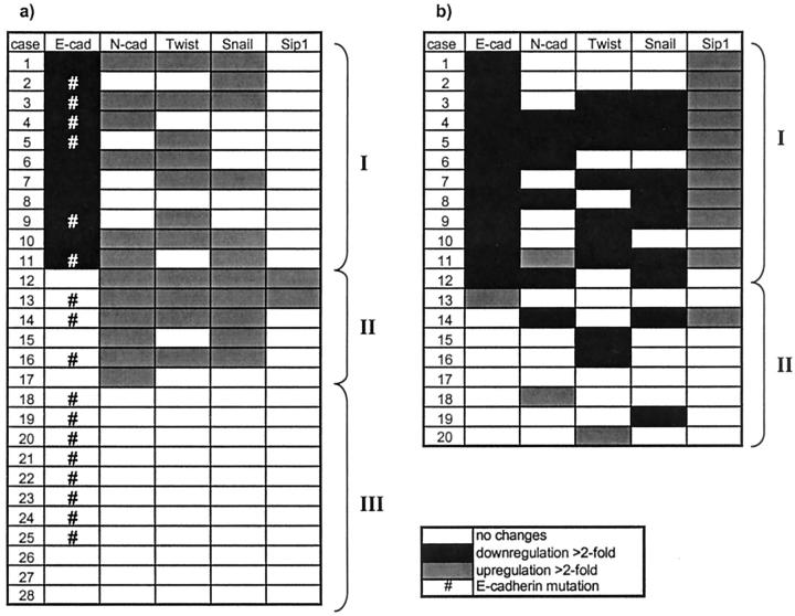
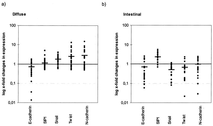
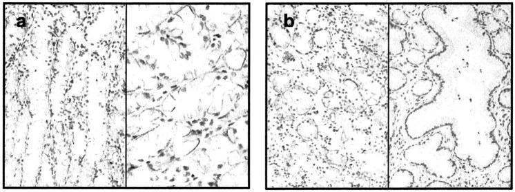
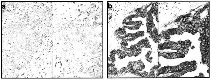
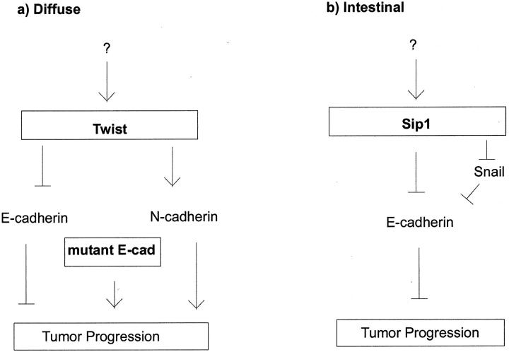
Epithelial-mesenchymal transition (EMT) involving down-regulation of E-cadherin is thought to play a fundamental role during early steps of invasion and metastasis of carcinoma cells. The aim of our study was to elucidate the role of EMT regulators Snail, SIP1 (both are direct repressors of E-cadherin), and Twist (an activator of N-cadherin during Drosophila embryogenesis), in primary human gastric cancers. Expression of Snail, SIP1, and Twist was analyzed in 48 gastric carcinomas by real-time quantitative RT-PCR in paraffin-embedded and formalin-fixed tissues. The changes of expression levels of these genes in malignant tissues compared to matched non-tumorous tissues were correlated with the expression of E- and N-cadherin. From 28 diffuse-type gastric carcinomas analyzed reduced E-cadherin expression was detected in 11 (39%) cases compared to non-tumorous tissues. Up-regulated Snail could be found in 6 cases with reduced or negative E-cadherin expression. However, there was no correlation to increased SIP1 expression. Interestingly, we could detect abnormal expression of N-cadherin mRNA in 6 cases, which was correlated with Twist overexpression in 4 cases. From 20 intestinal-type gastric cancer samples reduced E-cadherin expression was found in 12 (60%) cases, which was correlated to up-regulation of SIP1, since 10 of these 12 cases showed elevated mRNA levels, whereas Snail, Twist, and N-cadherin were not up-regulated. We present the first study investigating the role of EMT regulators in human gastric cancer and provide evidence that an increase in Snail mRNA expression is associated with down-regulation of E-cadherin in diffuse-type gastric cancer. We detected abnormally positive or increased N-cadherin mRNA levels in the same tumors, probably due to overexpression of Twist. SIP1 overexpression could not be linked to down-regulated E-cadherin in diffuse-type tumors, but was found to be involved in the pathogenesis of intestinal-type gastric carcinoma. We conclude that EMT regulators play different roles in gastric carcinogenesis depending on the histological subtype.


Figures

Figure 1.
Figure 1.
Matrix of EMT regulator expression analyzed with QRT-PCR in malignant tissues compared to non-tumorous tissue in diffuse type gastric cancer (a) and in intestinal type gastric cancer (b). Cases were arranged from top to bottom, beginning with the highest down-regulation of E-cadherin mRNA, to show the relationship to N-cadherin and EMT regulator expression.
Figure 2.
Figure 2.
Changes in expression levels comparing tumor to non-tumorous samples logarithmically scaled. Horizontal bars (mean of changes in expression) represent a typical expression pattern of EMT regulators in relation to E-cadherin down-regulation. a: Diffuse type tumors show down-regulation of E-cadherin which is accompanied by up-regulation of Snail, Twist, and N-cadherin, but SIP1 is nearly equally expressed. b: The molecular profile of intestinal type gastric cancer shows that down-regulation of E-cadherin is accompanied by increases of SIP1, whereas Snail, Twist, and N-cadherin are even down-regulated, which is nearly the reverse expression pattern seen in diffuse type tumors (except for E-cadherin).
Figure 3.
Figure 3.
Immunoblot analysis of N-cadherin (a) and E-cadherin expression detected by ECL (b). 15 μg of total protein from MDA MB-435s cell lysates transfected with normal E-cadherin cDNA were used for the analysis. Molecular weights of the protein are indicated.
Figure 4.
Figure 4.
Immunohistochemistry of diffuse-type gastric carcinoma with a specific monoclonal N-cadherin antibody and counterstained with hematoxylin. a: Ganglion cells were used as positive control (magnifications: left, ×100; right, ×200). b: 20% tumor cells show membranous or weak cytoplasmic staining (case no. 11; magnifications: ×100 and ×200). c: No immunodetectable protein expression though abundant N-cadherin mRNA (case no. 4; magnifications: left, ×100; right, ×200).
Figure 5.
Figure 5.
Immunohistochemistry of non-tumorous gastric glands counterstained with hematoxylin. a: Unexpected positive N-cadherin immunoreaction in chief and parietal cells (magnifications: left, ×100; right, ×200). b: Comparison of deep gastric glands with foveolar glands, where N-cadherin immunoreactivity is seen in chief and parietal cells but not in foveolar epithelium (magnification, ×100).
Figure 6.
Figure 6.
Immunohistochemistry of intestinal-type gastric carcinomas with the specific monoclonal E-cadherin antibody and counterstained with hematoxylin. a: Tumor cells show low protein expression consistent with TaqMan results (magnifications: left, ×100; right, ×200). b: High expression of E-cadherin in tumor cells consistent with TaqMan results (magnifications: left, ×100; right, ×200).
Figure 7.
Figure 7.
Proposed model of E-cadherin dependent carcinogenesis pathways of diffuse type (a) and intestinal type gastric cancer (b). Based on our findings, we propose a key role for the E- to N-cadherin switch, beside loss of E-cadherin function through gene mutations, in diffuse type gastric carcinoma. In intestinal type gastric carcinoma, enhanced SIP1 expression may be involved in tumor progression due to E-cadherin repression.

Similar articles

Cited by

References

    1. Hirohashi S: Inactivation of the E-cadherin-mediated cell adhesion system in human cancers. Am J Pathol 1998, 153:333-339 - PMC - PubMed
    1. Hsu MY, Meier FE, Nesbit M, Hsu JY, Van Belle P, Elder DE, Herlyn M: E-cadherin expression in melanoma cells restores keratinocyte-mediated growth control and down-regulates expression of invasion-related adhesion receptors. Am J Pathol 2000, 156:1515-1525 - PMC - PubMed
    1. Cavallaro U, Christofori G: Cell adhesion in tumor invasion and metastasis: loss of the glue is not enough. Biochim Biophys Acta 2001, 1552:39-45 - PubMed
    1. Boyer B, Valles AM, Edme N: Induction and regulation of epithelial-mesenchymal transitions. Biochem Pharmacol 2000, 60:1091-1099 - PubMed
    1. Rashid MG, Sanda MG, Vallorosi CJ, Rios-Doria J, Rubin MA, Day ML: Post-translational truncation and inactivation of human E-cadherin distinguishes prostate cancer from matched normal prostate. Cancer Res 2001, 61:489-492 - PubMed

Publication types

MeSH terms