. 2002 Dec;76(23):11827-36.
doi: 10.1128/jvi.76.23.11827-11836.2002.

Capture and transfer of simian immunodeficiency virus by macaque dendritic cells is enhanced by DC-SIGN

Affiliations

Capture and transfer of simian immunodeficiency virus by macaque dendritic cells is enhanced by DC-SIGN

Monica T Yu Kimata et al. J Virol. 2002 Dec.

Abstract

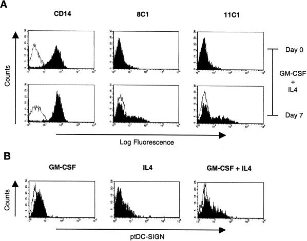
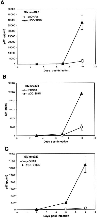
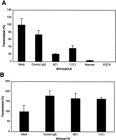
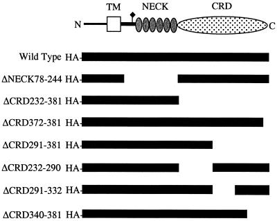
Dendritic cells (DCs) are among the first cells encountered by human and simian immunodeficiency virus (HIV and SIV) following mucosal infection. Because these cells efficiently capture and transmit virus to T cells, they may play a major role in mediating HIV and SIV infection. Recently, a C-type lectin protein present on DCs, DC-specific ICAM-3-grabbing nonintegrin (DC-SIGN), was shown to efficiently bind and present HIV and SIV to CD4(+), coreceptor-positive cells in trans. However, the significance of DC-SIGN for virus transmission and pathogenesis in vivo remains unclear. Because SIV infection of macaques may represent the best model to study the importance of DC-SIGN in HIV infection, we cloned and characterized pig-tailed macaque DC-SIGN and generated monoclonal antibodies (MAbs) against it. We demonstrate that, like human DC-SIGN, pig-tailed macaque DC-SIGN (ptDC-SIGN) is expressed on DCs and macrophages but not on monocytes, T cells, or B cells. Moderate levels of ptDC-SIGN expression were detected on the surface of DCs, and low-level expression was found on macrophages. Additionally, we show that ptDC-SIGN efficiently binds and transmits replication-competent SIVmne variants to CD4(+), coreceptor-positive cells. Moreover, transmission of virus between pig-tailed macaque DCs and CD4(+) T cells is largely ptDC-SIGN dependent. Interestingly, MAbs directed against ptDC-SIGN vary in the capacity to block transmission of different SIVmne variants. These data demonstrate that ptDC-SIGN plays a central role in transmitting virus from macaque DCs to T cells, and they suggest that SIVmne variants may differ in their interactions with ptDC-SIGN. Thus, SIVmne infection of pig-tailed macaques may provide an opportunity to investigate the significance of DC-SIGN in primate lentiviral infections.


Figures

FIG. 1.
FIG. 1.
Reactivity of MAbs raised against ptDC-SIGN. 293T cells were transfected with either a control, ptDC-SIGN, or huDC-SIGN expression vector. Two days posttransfection, cells were harvested, bound with either anti-ptDC-SIGN MAb 8C1 or 11C1 (filled curves), or the appropriate isotypic control antibodies (open curves), and a goat-anti-mouse FITC-labeled secondary antibody. Cells were then analyzed by FACS.
FIG. 2.
FIG. 2.
ptDC-SIGN expression on CD14+ monocytes, macrophages, and dendritic cells. CD14+ monocytes were positively selected from pig-tailed macaque PBMCs and stained for either CD14 or ptDC-SIGN before and after 7 days of culture with GM-CSF and IL-4 (A) or after 7 days of culture with GM-CSF alone, IL-4 alone, or GM-CSF plus IL-4 (B). Cells stained with either anti-CD14 or anti-ptDC-SIGN are shown in the filled curves, and isotype controls are shown in the open curves. Similar results were observed in two independent experiments using cells from two different macaques.
FIG. 3.
FIG. 3.
Enhancement of SIV transmission by ptDC-SIGN. 293T cells were transfected with either a control (pcDNA3) or ptDC-SIGN expression vector (pcDNA-ptDC-SIGN). Cells were incubated with SIVmneCL8 (A), SIVmne170 (B), or SIVmne027 (C), washed, and cultured with CEMx174 cells. Virus production was monitored by measuring p27gag antigen in the culture supernatants over a 7- to 10-day period. Results are representative of at least three independent experiments. The average p27gag antigen values ± the standard error of the mean are shown.
FIG. 4.
FIG. 4.
Interference of SIV capture and transmission by anti-ptDC-SIGN MAbs. 293T cells expressing ptDC-SIGN were mock treated or incubated for 20 min with 25 μg of control mouse IgG, 8C1, or 11C1 per ml prior to the addition of SIVmneCL8 (A) or SIVmne170 (B). Cells were also treated with mannan (20 μg/ml) or EGTA (5 mM) prior to the addition of SIVmneCL8. Following a 2-h incubation with virus, cells were washed and cultured with CEMx174 cells. Virus production was measured by assaying supernatants for p27gag antigen by ELISA over a 7-day period. Day 7 p27gag antigen values of the mock-treated cells were normalized to 100%. The amount of p27gag antigen production from each infection is shown relative to the amount produced from the mock-treated cells and represents the average ± the standard error of the mean. Data are representative of at least two independent experiments.
FIG. 5.
FIG. 5.
Capture and transmission of SIV by macaque DCs. Duplicate cultures of DCs were mock treated or incubated with 25 μg of control IgG, 8C1, or 11C1 per ml for 20 min prior to the addition of SIVmne027. Following a 3-h incubation with virus, the cells were washed and incubated with PHA-stimulated PBMCs and IL-2. Virus production was monitored by p27gag ELISA every 3 to 4 days postinfection. The average p27gag antigen values ± the standard error of the mean are shown. The experiments shown in panels A and B were performed using cells isolated from two different macaques.
FIG. 6.
FIG. 6.
Inhibition of huDC-SIGN-dependent HIV-1 capture and transfer by the anti-ptDC-SIGN MAb. THP-1/huDC-SIGN cells were mock treated or incubated with 50 μg of anti-ptDC-SIGN MAb 11C1 (A) or 8C1 (B) per ml prior to incubation with an HIV-luc reporter virus pseudotyped with either the HIV-1ADA or HIV-1JRFL envelope. Following the incubation with virus, cells were washed and cultured with HOS-CD4-CCR5 cells. Viral infection was determined by measuring luciferase activity in the target cells. THP-1/DC-SIGN cells were used for background virus capture and transfer. Relative luciferase activity ± the standard error of the mean is shown.
FIG. 7.
FIG. 7.
Deletion constructs of ptDC-SIGN. The portion of ptDC-SIGN present in each deletion mutant is shown relative to that in the wild-type clone. Also shown is a schematic diagram of the ptDC-SIGN molecule.
FIG. 8.
FIG. 8.
Reactivity of anti-DC-SIGN MAbs with deletion mutants of ptDC-SIGN. 293T cells were transfected with vectors expressing the wild-type ptDC-SIGN, a deletion of the complete CRD (ΔCRD232-381), or deletion of the complete neck domain (ΔNECK78-244). Cell expressing each construct were stained with the 8C1, 11C1, or anti-neck antibody DC4 (black curves) or stained with isotypic control antibodies (open curves) and analyzed by FACS.

Similar articles

Cited by

References

    1. Adema, G. J., F. Hartgers, R. Verstraten, E. de Vries, G. Marland, S. Menon, J. Foster, Y. Xu, P. Nooyen, T. McClanahan, K. B. Bacon, and C. G. Figdor. 1997. A dendritic-cell-derived C-C chemokine that preferentially attracts naive T cells. Nature 387:713-717. - PubMed
    1. Baribaud, F., S. Pohlmann, T. Sparwasser, M. T. Kimata, Y. K. Choi, B. S. Haggarty, N. Ahmad, T. Macfarlan, T. G. Edwards, G. J. Leslie, J. Arnason, T. A. Reinhart, J. T. Kimata, D. R. Littman, J. A. Hoxie, and R. W. Doms. 2001. Functional and antigenic characterization of human, rhesus macaque, pig-tailed macaque, and murine DC-SIGN. J. Virol. 75:10281-10289. - PMC - PubMed
    1. Bell, D., J. W. Young, and J. Banchereau. 1999. Dendritic cells. Adv. Immunol. 72:255-324. - PubMed
    1. Bukrinsky, M. I., T. L. Stanwick, and M. Stevenson. 1991. Quiescent T lymphocytes as an inducible virus reservoir in HIV1 infection. Science 254:423-427. - PMC - PubMed
    1. Cameron, P., M. Pope, A. Granelli-Piperno, and R. M. Steinman. 1996. Dendritic cells and the replication of HIV-1. J. Leukoc. Biol. 59:158-171. - PubMed

Publication types

MeSH terms

Substances