. 2002 Dec;76(23):11866-79.
doi: 10.1128/jvi.76.23.11866-11879.2002.

Mapping of functional regions in the amino-terminal portion of the herpes simplex virus ICP27 regulatory protein: importance of the leucine-rich nuclear export signal and RGG Box RNA-binding domain

Affiliations

Mapping of functional regions in the amino-terminal portion of the herpes simplex virus ICP27 regulatory protein: importance of the leucine-rich nuclear export signal and RGG Box RNA-binding domain

Joy Lengyel et al. J Virol. 2002 Dec.

Abstract

Infected-cell protein 27 (ICP27) is an essential herpes simplex virus type 1 (HSV-1) regulatory protein that activates a subset of viral delayed-early and late genes, at least in part through posttranscriptional mechanisms. Previous studies have shown that the amino (N)-terminal half of the protein contains important functional regions, including a leucine-rich nuclear export signal (NES). However, to date, the phenotype of an HSV-1 ICP27 NES mutant has not been reported. In this study, we engineered and characterized dLeu, an HSV-1 deletion mutant that specifically lacks ICP27's NES (amino acids 6 to 19). The phenotype of dLeu was analyzed alongside those of eight other ICP27 N-terminal deletion mutants. We found that in Vero cells, dLeu displays modest defects in viral gene expression and an approximately 100-fold reduction in the production of viral progeny. Unlike wild-type (WT) ICP27, which exhibits a cytoplasmic distribution in addition to its predominant nuclear localization, dLeu ICP27 is highly restricted to the cell nucleus. This strongly suggests that the N-terminal leucine-rich sequence functions as an NES during viral infection. Our analysis of dLeu and the other mutants has enabled us to genetically define the regions in the N-terminal 200 residues of ICP27 which are required for efficient viral growth in Vero cells. Only two regions appear to be important: (i) the leucine-rich NES and (ii) the RGG box RNA-binding domain, encoded by residues 139 to 153. A virus lacking the RGG box-encoding sequence, d4-5, has a phenotype similar to that of dLeu in that it displays modest defects in viral gene expression and grows poorly. Interestingly, deletion of both the NES and RGG box, as well as the sequences in between, is lethal. The resulting virus, d1-5, displays severe defects in viral gene expression and DNA synthesis and is unable to produce significant amounts of infectious progeny. Therefore, the N-terminal portion of ICP27 contains at least two functional domains which collectively are absolutely essential for viral infection.


Figures

FIG. 1.
FIG. 1.
HSV-1 ICP27 N-terminal mutants. (A) The bar at the top represents the ICP27 polypeptide, highlighting known motifs and functional sequences. Below the bar, numbers 1 to 7 refer to sites of engineered XhoI restriction sites (34) which were used to construct many of the deletion mutations. The regions of ICP27 that harbor the epitopes for mouse MAbs H1119 and H1113 are indicated. Also indicated are the leucine-rich NES, acidic region, NLS 1, NLS 2, RGG box RNA-binding domain, and regions highly conserved in the ICP27 homologs of other herpesviruses (CR1 to -3). The solid horizontal lines below the bar indicate the ICP27 protein sequences present in the various N-terminal deletion mutants; dashed lines indicate in-frame deletions. (B) Comparison of amino acid sequences deleted in dLeu, d1-2, and dAc. The N-terminal 70 residues (in single-letter amino acid code) of ICP27 from strain KOS1.1 are shown, along with the location of ICP27's N-terminal leucine-rich NES. The extents of the deletions in dLeu, d1-2, and dAc are indicated by lines.
FIG. 2.
FIG. 2.
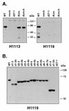
Immunoblot analysis of N-terminal deletion mutants. (A) Analysis of dLeu. Vero cells were mock infected or infected with the indicated viruses at an MOI of 10. Protein extracts were prepared at 5 hpi, and immunoblotting was carried out using MAbs H1113 and H1119, as indicated. The migration of marker proteins is shown. (B) Analysis of additional N-terminal mutants. The experiment was performed as for panel A, except that infections were carried out at an MOI of 1 and protein extracts were prepared at 16 hpi. Immunoblotting was performed with the H1119 MAb.
FIG. 3.
FIG. 3.
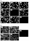
Cellular localization of mutant ICP27 polypeptides. Vero cells growing on coverslips were infected at an MOI of 10 with the viruses indicated. At 6 hpi, the cells were fixed and processed for immunofluorescence using anti-ICP27 MAb H1113 (A) or H1119 (B). For each panel, staining, image capture, and image manipulation were done in parallel so that the localization patterns of the different mutant proteins could be directly compared.
FIG. 4.
FIG. 4.
Viral yields in infected Vero and V27 cells. (A) Single-cycle yield assays. Replicate cultures of approximately 2 × 10⁶ Vero or V27 cells were infected in duplicate at an MOI of 1 with WT HSV-1 or various ICP27 mutants. After 24 h, the infections were terminated by freezing. Virus was released from the cells by three cycles of freeze-thawing, and the total infectious virus in the lysate was determining by a plaque assay on V27 cells. The gray and hatched bars represent the mean yields in V27 and Vero cells, respectively. Error bars indicate the values of the duplicate samples. (B) Growth curves. Cultures of approximately 2 × 10⁶ Vero cells were infected in duplicate at an MOI of 1 with WT HSV-1 or various ICP27 mutants. Infections were terminated at multiple time points by freezing, and virus yields were determined as for panel A. The values shown represent the mean yields of duplicate infections.
FIG. 5.
FIG. 5.
Viral DNA replication by ICP27 mutants. Replicate cultures of Vero cells were mock infected or infected with WT HSV-1 or ICP27 mutants at an MOI of 10. The cells were labeled with [3H]thymidine from 7 to 13 hpi. After purification of total cellular DNA, equal amounts (2.5 μg) were digested with BamHI and electrophoresed on a 1% agarose gel. The gel was treated with a fluorography enhancing agent, dried, and exposed to X-ray film. An autoradiograph is shown. The lines to the right indicate the migration of viral BamHI restriction fragments.
FIG. 6.
FIG. 6.
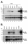
Expression of UL42 and gC mRNAs in mutant-infected Vero cells. (A) Expression of UL42 mRNA. Replicate cultures of Vero cells were mock infected or infected at an MOI of 10 with the viruses indicated. Where indicated, PAA was included in the culture medium at a concentration of 400 μg/ml to inhibit viral DNA replication. At 6 hpi, total RNA was isolated, and equal amounts were analyzed by Northern blotting with a UL42 probe (top panel). The blot was then stripped and reprobed with an ICP8 mRNA-specific probe (bottom panel). (B) Expression of gC mRNA. Replicate cultures of Vero cells were mock infected or infected with the viruses indicated at an MOI of 10. At 9.5 hpi, total RNA was isolated and equal amounts were analyzed by Northern blotting with a gC gene-specific probe (top panel). After stripping of the blot, a second hybridization was performed using an ICP8-specific probe (bottom panel).
FIG. 7.
FIG. 7.
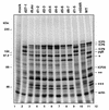
Protein synthesis in ICP27 mutant-infected Vero cells. Replicate cultures of Vero cells were mock infected or infected at an MOI of 10 by the viruses indicated. At 9 to 9.5 hpi, the cultures were labeled with [35S]methionine-cysteine and total cell protein samples were prepared. The proteins were separated by SDS-polyacrylamide gel electrophoresis and analyzed by autoradiography. The positions at which molecular mass markers migrated are indicated at the left, and the positions of known viral proteins are indicated at the right. The positions of unidentified infection-specific proteins discussed in the text are indicated at the right by asterisks.
FIG. 8.
FIG. 8.
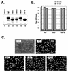
Deletion of the acidic region between residues 21 and 63 does not affect viral growth or ICP27 localization. (A) Immunoblot analysis of dAc ICP27. Vero cells were mock infected or infected with the viruses indicated at an MOI of 10. Protein extracts were prepared at 5 hpi, and immunoblotting was carried out with MAb H1113. (B) Plaquing ability of dAc. Stocks of WT HSV-1 or dAc isolates were titrated by plaque assay on Vero cells (gray bars) or V27 cells (hatched bars). (C) Localization of ICP27 in dAc-infected cells. Vero cells were mock infected or infected with the viruses indicated. The cells were fixed at 6 hpi and processed for immunofluorescence with the H1113 MAb.

Similar articles

Cited by

References

    1. Aubert, M., S. A. Rice, and J. A. Blaho. 2001. Accumulation of herpes simplex virus type 1 early and leaky-late proteins correlates with apoptosis prevention in infected human HEp-2 cells. J. Virol. 75:1013-1030. - PMC - PubMed
    1. Blaho, J. A., C. Mitchell, and B. Roizman. 1994. An amino acid sequence shared by the herpes simplex virus 1 alpha regulatory proteins 0, 4, 22, and 27 predicts the nucleotidylylation of the UL21, UL31, UL47, and UL49 gene products. J. Biol. Chem. 269:17401-17410. - PubMed
    1. Blaho, J. A., C. Mitchell, and B. Roizman. 1993. Guanylylation and adenylylation of the alpha regulatory proteins of herpes simplex virus require a viral beta or gamma function. J. Virol. 67:3891-3900. - PMC - PubMed
    1. Brown, C. R., M. S. Nakamura, J. D. Mosca, G. S. Hayward, S. E. Straus, and L. P. Perera. 1995. Herpes simplex virus trans-regulatory protein ICP27 stabilizes and binds to 3′ ends of labile mRNA. J. Virol. 69:7187-7195. - PMC - PubMed
    1. Bryant, H. E., D. A. Matthews, S. Wadd, J. E. Scott, J. Kean, S. Graham, W. C. Russell, and J. B. Clements. 2000. Interaction between herpes simplex virus type 1 IE63 protein and cellular protein p32. J. Virol. 74:11322-11328. - PMC - PubMed

Publication types

MeSH terms

LinkOut - more resources